ACTA
PHARMACEUTICA
SCIENCIA
International Journal in Pharmaceutical Sciences, Published Quarterly
ISSN: 2636-8552
e-ISSN: 1307-2080,
Volume: 57, No: 2, 2019
Formerly: Eczacılık Bülteni
Acta Pharmaceutica Turcica

Fatih Demirci (Anadolu University, Turkey)
Hakan Göker (Ankara University, Turkey)
Hanefi Özbek
(İstanbul Medipol University, Turkey)
Hayati Çelik (Yeditepe University, Turkey)
İhsan Çalış (Near East University, Cyprus)
Julide Akbuğa (Altınbaş University, Turkey)
Kenneth A. Jacobson
(National Institutes of Health, USA)
Leyla Yurttaş (Anadolu University, Turkey)
Mahmud Miski (İstanbul University, Turkey)
Mesut Sancar (Marmara University, Turkey)
Murat Duran
(Eskişehir Osmangazi University, Turkey)
Nesrin Emekli
(İstanbul Medipol University, Turkey)
Nilay Aksoy (Altınbaş University, Turkey)
Nurşen Başaran (Hacettepe University, Turkey)
Özgen Özer (Ege University, Turkey)
Roberta Ciccocioppo
(University of Camerino, Italy)
Selma Saraç Tarhan
(Hacettepe University, Turkey)
Semra Şardaş (İstinye University, Turkey)
Sevda Süzgeç Selçuk
(İstanbul University, Turkey)
Stefano Constanzi (American University, USA)
Süreyya Ölgen (Biruni University, Turkey)
Şule Apikoğlu Rabuş
(Marmara University, Turkey)
Tuncer Değim (Biruni University, Turkey)
Yıldız Özsoy (İstanbul University, Turkey)
Yusuf Öztürk (Anadolu University, Turkey)
Printing Office
Forart Basımevi
Ziya Gökalp Mah.
Süleyman Demirel Bulvarı
Simpaş İş Modern Zemin Kat A 17-18
İkitelli - İstanbul
Tel: (0212) 501 82 20
Editor
Şeref Demirayak
Associate Editors
Gülden Zehra Omurtag
Barkın Berk
Coordinators
M. Eşref Tatlıpınar
Gökberk Karabay
Language Editor
Recep Murat Nurlu
M. Eşref Tatlıpınar
Neda Taner
Biostatistics Editor
Pakize Yiğit
Graphic Design
Sertan Vural
Levent Karabağlı
Address
İstanbul Medipol Üniversitesi
Kavacık Güney Kampüsü
Göztepe Mah. Atatürk Cad.
No: 40 34810 Beykoz/İSTANBUL
Tel: 0216 681 51 00
E-mail
Web site
http://www.actapharmsci.com
Editorial Board
Sabahattin Aydın
(Istanbul Medipol University, Turkey)
Ahmet Aydın (Yeditepe University, Turkey)
Ahmet Çağrı Karaburun (Anadolu University, Turkey)
Aristidis Tsatsakis (University of Crete, Greece)
Ayfer Beceren (Marmara University, Turkey)
Dilek Ak (Anadolu University, Turkey)
Ebrahim Razzazi-Fazeli
(University of Veterinary Medicine, Vienna)
Erem Memişoğlu Bilensoy
(Hacettepe University, Turkey)
Fatma Tosun
(İstanbul Medipol University, Turkey)
ACTA
PHARMACEUTICA
SCIENCIA
International Journal in Pharmaceutical Sciences
is Published Quarterly
ISSN: 2636-8552
e-ISSN: 1307-2080,
Volume: 57, No: 2, 2019
Formerly: Eczacılık Bülteni/Acta Pharmaceutica Turcica
Founded in 1953 by Kasım Cemal Güven
Contents
Aims and Scope of Acta Pharmaceutica Sciencia
Şeref Demirayak ......................................................113
Instructions for Authors ...............................................114
Original articles ...................................................... 128
Synthesis, Characterization and Evaluation of the Anti-cancer Activity of Silver
Nanoparticles by Natural Organic Compounds Extracted from Cyperus sp.
rhizomes
Rasim Farraj Muslim, Mustafa Nadhim Owaid ............................... 129
Anti-inflammatory and Analgesic Potential of Acetone Leaf Extract of Combretum
Sordidum and its Fractions
Babatunde Samuel, Olayinka Oridupa, Fisayo Gbadegesin ....................147
Combination of Cumulative Area Pre-Processing and Partial Least Squares for
Handling Intensely Overlapping Binary and Ternary Drug Systems
Yahya ِAl-Degs, Amjad El-Sheikh, Eman Abu Saaleek, Reema Omeir, Musab Al-
Ghodran ............................................................161
Chemical Composition and Comparative Antibacterial Properties of Basil
Essential Oil against Clinical and Standard Strains of Campylobacter spp.
Aysegul Mutlu-Ingok, Burcu Firtin, Funda Karbancioglu-Guler .................. 183
In vitro Antimicrobial and Antioxidant Evaluation of Melampyrum Arvense Var.
Elatius L. and Sedum Spurium M. Bieb. Extracts
Ayşe Esra Karadağ, Fatma Tosun ........................................193
Preparation and In vitro Characterization of a Fluconazole Loaded Chitosan
Particulate System
Gülsel Yurtdaş-Kırımlıoğlu, Yenilmez Evrim, Başaran Ebru, Yazan Yasemin
Review articles ...................................................... 216
Pharmaceutical Properties of Marine Polyphenols:
An Overview
Thanh Sang Vo, Dai Hung Ngo, Se-Kwon Kim ...............................217

113
Acta Pharmaceutica Sciencia. Vol. 57 No. 2, 2019
Aims and Scope of Acta Pharmaceutica Sciencia
Acta Pharmaceutica Sciencia is a continuation of the former “Eczacılık Bülteni”
which was first published in 1953 by Prof. Dr. Kasım Cemal Güven’s editorship.
At that time, “Eczacılık Bülteni” hosted scientific papers from the School of Med-
icine-Pharmacy at Istanbul University, Turkey.
In 1984, the name of the journal was changed to “Acta Pharmaceutica Turcica”
and it became a journal for national and international manuscripts, in all fields
of pharmaceutical sciences in both English and Turkish. (1984-1995, edited
by Prof. Dr. Kasım Cemal Güven, 1995-2001, edited by Prof. Dr. Erden Güler,
2002-2011, edited by Prof. Dr. Kasım Cemal Güven)
Since 2006, the journal has been published only in English with the name, “Acta
Pharmaceutica Sciencia” which represents internationally accepted high-level
scientific standards. The journal has been published quarterly except for an in-
terval from 2002 to 2009 in which its issues were released at intervals of four
months. The publication was also temporarily discontinued at the end of 2011
but since 2016, Acta Pharmaceutica Sciencia has continued publication with the
reestablished Editorial Board and also with the support of you as precious sci-
entists.
Yours Faithfully
Prof. Dr. Şeref DEMİRAYAK
Editor

114
Acta Pharmaceutica Sciencia. Vol. 57 No. 2, 2019
INSTRUCTIONS FOR AUTHORS
1. Scope and Editorial Policy
1.1. Scope of the Journal
Acta Pharmaceutica Sciencia (Acta Pharm. Sci.), formerly known as Bulletin of
Pharmacy and Acta Pharmaceutica Turcica is a peer-reviewed scientific jour-
nal publishing current research and reviews covering all fields of pharmaceu-
tical sciences since 1953.
The original studies accepted for publication must be unpublished work and
should contain data that have not been published elsewhere as a whole or a
part. The reviews must provide critical evaluation of the state of knowledge
related with the subject.
All manuscripts has to be written in clear and concise English.
Starting from 2016, the journal will be issued quarterly both in paper and on-
line formates also publish special issues for national or international scientific
meetings and activities in the coverage field.
1.2 Manuscript Categories
Manuscripts can be submitted as Research Articles and Reviews.
1.2.1 Research Articles are definitive accounts of significant, original studies.
They are expected to present important new data or provide a fresh approach
to an established subject.
1.2.2 Reviews integrate, correlate, and evaluate results from published litera-
ture on a particular subject. They expected to report new and up to date expe-
rimental findings. They have to have a well-defined theme, are usually critical,
and may present novel theoretical interpretations. Up to date experimental
procedures may be included. Reviews are usually submitted at the invitation
of the Editors. However, experts are welcome to contact the Editors to ensure
that a topic is suitable. Approval is recommended prior to submission.
1.3 Prior Publication
Authors should submit only original work that has not been previously publis-
hed and is not under consideration for publication elsewhere. Academic the-
ses, including those on the Web or at a college Web site, are not considered to
be prior publication.
1.4 Patents and Intellectual Property
Authors need to resolve all patent and intellectual property issues. Acceptance

115
Acta Pharmaceutica Sciencia. Vol. 57 No. 2, 2019
and publication will not be delayed for pending or unresolved issues of this
type. Note that Accepted manuscripts and online manuscripts are considered
as published documents.
1.5 Professional Ethics
Editors, reviewers, and authors are expected to adhere to internationally ac-
cepted criteria’s for scientific publishing.
1.5.1 Author Consent. Submitting authors are reminded that consent of all
coauthors must be obtained prior to submission of manuscripts. If an author
is removed after submission, the submitting author must have the removed
author consent to the change by e-mail or faxed letter to the assigned Editor.
1.5.2. Plagiarism. Manuscripts must be original with respect to concept, con-
tent, and writing. It is not appropriate for an author to reuse wording from other
publications, including one›s own previous publications, whether or not that
publication is cited. Suspected plagiarism should be reported immediately to
the editorial oice. Report should specifically indicate the plagiarized material
within the manuscripts. Acta Pharmaceutica Sciencia uses iThenticate or Turni-
tin software to screen submitted manuscripts for similarity to published materi-
al. Note that your manuscript may be screened during the submission process.
1.5.3. Use of Human or Animal Subjects. For research involving biolo-
gical samples obtained from animals or human subjects, editors reserve the
right to request additional information from authors. Studies submitted for
publication approval must present evidence that the described experimental
activities have undergone local institutional review assessing safety and hu-
mane usage of study subject animals. In the case of human subjects authors
must also provide a statement that study samples were obtained through the
informed consent of the donors, or in lieu of that evidence, by the authority
of the institutional board that licensed the use of such material. Authors are
requested to declare the identification or case number of institution approval
as well as the name of the licensing committee in a statement placed in the
section describing the studies’ Material and Methods.
1.6 Issue Frequency
The Journal publishes 4 issues per year.
2. Preparing the Manuscript
2.1 General Considerations
Manuscripts should be kept to a minimum length. Authors should write in cle-

116
Acta Pharmaceutica Sciencia. Vol. 57 No. 2, 2019
ar, concise English, employing an editing service if necessary. For professional
assistance with improving the English, figures, or formatting in the manuscript
before submission please contact to editorial oice by e-mail for suggestions.
The responsibility for all aspects of manuscript preparation rests with the aut-
hors. Extensive changes or rewriting of the manuscript will not be undertaken
by the Editors. A standard list of Abbreviations, Acronyms and Symbols is in
section 5.
It is best to use the fonts “Times” and “Symbol.” Other fonts, particularly those
that do not come bundled with the system software, may not translate pro-
perly. Ensure that all special characters (e.g., Greek characters, math symbols)
are present in the body of the text as characters and not as graphic represen-
tations. Be sure that all characters are correctly represented throughout the
manuscript—e.g., 1 (one) and l (letter l), 0 (zero) and O (letter o).
All text (including the title page, abstract, all sections of the body of the paper,
figure captions, scheme or chart titles, and footnotes and references) and tab-
les should be in one file. Graphics may be included with the text or uploaded as
separate files. Manuscripts that do not adhere to the guidelines may be retur-
ned to authors for correction.
2.1.1 Articles of all kind. Use page size A4. Vertically orient all pages. Ar-
ticles of all kind must be double-spaced including text, references, tables, and
legends. This applies to figures, schemes, and tables as well as text. They do not
have page limitations but should be kept to a minimum length. The experimen-
tal procedures for all of experimental steps must be clearly and fully included
in the experimental section of the manuscripts.
2.1.2 Nomenclature. It is the responsibility of the authors to provide cor-
rect nomenclature. It is acceptable to use semisynthetic or generic names for
certain specialized classes of compounds, such as steroids, peptides, carbohy-
drates, etc. In such a case, the name should conform to the generally accepted
nomenclature conventions for the compound class. Chemical names for drugs
are preferred. If these are not practical, generic names, or names approved by
the World Health Organization, may be used.
Authors may find the following sources useful for recommended nomenclatu-
re:
· The ACS Style Guide; Coghill, A. M., Garson, L. R., Eds.; American Chemical
Society: Washington DC, 2006.
· Enzyme Nomenclature; Webb, E. C., Ed.; Academic Press: Orlando, 1992.

117
Acta Pharmaceutica Sciencia. Vol. 57 No. 2, 2019
· IUPHAR database of receptors and ion channels (http://www.guidetophar-
macology.org/).
2.1.3 Compound Code Numbers. Code numbers (including peptides) as-
signed to a compound may be used as follows:
· Once in the manuscript title, when placed in parentheses AFTER the chemical
or descriptive name.
· Once in the abstract.
· Once in the text (includes legends) and once to label a structure. Code num-
bers in the text must correspond to structures or, if used only once, the chemi-
cal name must be provided before the parenthesized code number, e.g., “che-
mical name (JEM-398).” If appearing a second time in the text, a bold Arabic
number must be assigned on first usage, followed by the parenthesized code
number, e.g., “1 (JEM-398).” Subsequently, only the bold Arabic number may
be used. All code numbers in the text must have a citation to a publication or a
patent on first appearance.
Compounds widely employed as research tools and recognized primarily by
code numbers may be designated in the manuscript by code numbers without
the above restrictions. Their chemical name or structure should be provided
as above. Editors have the discretion of determining which code numbers are
considered widely employed.
2.1.4 Trademark Names. Trademark names for reagents or drugs must be
used only in the experimental section. Do not use trademark or service mark
symbols.
2.1.5 Interference Compounds. Active compounds from any source must
be examined for known classes of assay interference compounds and this
analysis must be provided in the General Experimental section. Many of these
compounds have been classified as Pan Assay Interference Compounds (PA-
INS; see Baell & Holloway, J. Med. Chem. 2010, 53, 2719-2740). These compo-
unds shown to display misleading assay readouts by a variety of mechanisms
by forming reactive compounds. Provide firm experimental evidence in at least
two dierent assays that reported compounds with potential PAINS liability
are specifically active and their apparent activity is not an artifact.
2.2 Manuscript Organization
2.2.1 Title Page. Title: The title of the manuscript should reect the pur-
poses and findings of the work in order to provide maximum information in a

118
Acta Pharmaceutica Sciencia. Vol. 57 No. 2, 2019
computerized title search. Minimal use of nonfunctional words is encouraged.
Only commonly employed abbreviations (e.g., DNA, RNA, ATP) are acceptab-
le. Code numbers for compounds may be used in a manuscript title when pla-
ced in parentheses AFTER the chemical or descriptive name.
Authors’ Names and Ailiations: The authors’ full first names, middle initials,
last names, and ailiations with addresses at time of work completion should
be listed below the title. The name of the corresponding author should be mar-
ked with an asterisk (*).
2.2.2 Abstract and keywords. Articles of all types must have an abstract
following the title page. The maximum length of the Abstract should be 150
words, organized in a findings-oriented format in which the most important
results and conclusions are summarized. Code numbers may be used once in
the abstract.
After the abstract, a section of Keywords not more than five has to be given.
Be aware that the keywords, chosen according to the general concept, are very
significant during searching and indexing of the manuscripts.
2.2.3 Introduction. The rationale and objectives of the research should be
discussed in this section. The background material should be brief and rele-
vant to the research described.
2.2.4. Methodology. Materials, synthetic, biological, demographic, statisti-
cal or experimental methods of the research should be given detailed in this
section. The authors are free to subdivide this section in the logical ow of the
study. For the experimental sections, authors should be as concise as possible
in experimental descriptions. General reaction, isolation, preparation conditi-
ons should be given only once. The title of an experiment should include the
chemical name and a bold Arabic identifier number; subsequently, only the
bold Arabic number should be used. Experiments should be listed in numeri-
cal order. Molar equivalents of all reactants and percentage yields of products
should be included. A general introductory section should include general pro-
cedures, standard techniques, and instruments employed (e.g., determination
of purity, chromatography, NMR spectra, mass spectra, names of equipment)
in the synthesis and characterization of compounds, isolates and preparations
described subsequently in this section. Special attention should be called to
hazardous reactions or toxic compounds. Provide analysis for known classes of
assay interference compounds.
The preferred forms for some of the more commonly used abbrevations are
mp, bp, ºC, K, min, h, mL, μL, g, mg, μg, cm, mm, nm, mol, mmol, μmol, ppm,

119
Acta Pharmaceutica Sciencia. Vol. 57 No. 2, 2019
TLC, GC, NMR, UV, and IR. Units are abbreviated in table column heads and
when used with numbers, not otherwise. (See section 4 for more abbreviations)
2.2.5 Results and Discussion. This section could include preparation, iso-
lation, synthetic schemes and tables of biological and statistical data. The dis-
cussions should be descriptive. Authors should discuss the analysis of the data
together with the significance of results and conclusions. An optional conclusi-
ons section is not required.
2.2.6 Ancillary Information. Include pertinent information in the order
listed immediately before the references.
PDB ID Codes: Include the PDB ID codes with assigned compound Arabic
number. Include the statement “Authors will release the atomic coordinates
and experimental data upon article publication.”
Homology Models: Include the PDB ID codes with assigned compound Arabic
number. Include the statement “Authors will release the atomic coordinates
upon article publication.”
Corresponding Author Information: Provide telephone numbers and email
addresses for each of the designated corresponding authors.
Present/Current Author Addresses: Provide information for authors whose
ailiations or addresses have changed.
Author Contributions: Include statement such as «These authors contributed
equally.»
Acknowledgment: Authors may acknowledge people, organizations, and fi-
nancial supporters in this section.
Abbreviations Used: Provide a list of nonstandard abbreviations and acronyms
used in the paper, e.g., YFP, yellow uorescent protein. Do not include compo-
und code numbers in this list. It is not necessary to include abbreviations and
acronyms from the Standard Abbreviations and Acronyms listed in section 4.
2.2.7 References and Notes. Number literature references and notes in one
consecutive series by order of mention in the text. Numbers in the text are non-
parenthesized superscripts. The accuracy of the references is the responsibility
of the author. List all authors; do not use et al. Provide inclusive page num-
bers. Titles may have capitalization of first word only (excluding, for example,
acronyms and trade names) or standard capitalization as shown below. The
chosen style should be used consistently throughout the references. Double-
space the references using the following format.

120
Acta Pharmaceutica Sciencia. Vol. 57 No. 2, 2019
· For journals: Rich, D. H.; Green, J.; Toth, M. V.; Marshall, G. R.; Kent, S. B.
H. Hydroxyethylamine Analogues of the p17/p24 Substrate Cleavage Site Are
Tight Binding Inhibitors of HIV Protease. J. Med. Chem. 1990, 33, 1285-1288.
· For online early access: Rubner, G.; Bensdorf, K.; Wellner, A.; Kircher, B.;
Bergemann, S.; Ott, I.; Gust, R. Synthesis and Biological Activities of Transiti-
on Metal Complexes Based on Acetylsalicylic Acid as Neo-Anticancer Agents. J.
Med. Chem. [Online early access]. DOI: 10.1021/jm101019j. Published Online:
September 21, 2010.
· For periodicals published in electronic format only: Author 1; Author 2; Aut-
hor 3; etc. Title of Article. Journal Abbreviation [Online] Year, Volume, Ar-
ticle Number or other identifying information.
· For monographs: Casy, A. F.; Parfitt, R. T. Opioid Analgesics; Plenum: New
York, 1986.
· For edited books: Rall, T. W.; Schleifer, L. S. Drugs Eective in the Therapy
of the Epilepsies. In The Pharmacological Basis of Therapeutics, 7th ed.; Gil-
man, A. G., Goodman, L. S., Rall, T. W., Murad, F., Eds.; Macmillan: New York,
1985; pp 446-472
List submitted manuscripts as “in press” only if formally accepted for publi-
cation. Manuscripts available on the Web with a DOI number are considered
published. For manuscripts not accepted, use “unpublished results” after the
names of authors. Incorporate notes in the correct numerical sequence with
the references. Footnotes are not used.
2.2.8 Tables. Tabulation of experimental results is encouraged when this le-
ads to more eective presentation or to more economical use of space. Tables
should be numbered consecutively in order of citation in the text with Arabic
numerals. Footnotes in tables should be given italic lowercase letter designa-
tions and cited in the tables as superscripts. The sequence of letters should
proceed by row rather than by column. If a reference is cited in both table and
text, insert a lettered footnote in the table to refer to the numbered reference
in the text. Each table must be provided with a descriptive title that, together
with column headings, should make the table self-explanatory. Titles and fo-
otnotes should be on the same page as the table. Tables may be created using
a word processor’s text mode or table format feature. The table format feature
is preferred. Ensure each data entry is in its own table cell. If the text mode is
used, separate columns with a single tab and use a return at the end of each
row. Tables may be inserted in the text where first mentioned or may be grou-
ped after the references.

121
Acta Pharmaceutica Sciencia. Vol. 57 No. 2, 2019
2.2.9 Figures, Schemes/Structures, and Charts. The use of illustrations
to convey or clarify information is encouraged. Structures should be produced
with the use of a drawing program such as ChemDraw. Authors using other
drawing packages should, in as far as possible, modify their program’s para-
meters so that they conform to ChemDraw preferences. Remove all color from
illustrations, except for those you would like published in color. Illustrations
may be inserted into the text where mentioned or may be consolidated at the
end of the manuscript. If consolidated, legends should be grouped on a separa-
te page(s). Include as part of the manuscript file.
To facilitate the publication process, please submit manuscript graphics using
the following guidelines:
1. The preferred submission procedure is to embed graphic files in a Word do-
cument. It may help to print the manuscript on a laser printer to ensure all
artwork is clear and legible.
2. Additional acceptable file formats are: TIFF, PDF, EPS (vector artwork)
or CDX (ChemDraw file). If submitting individual graphic files in addition to
them being embedded in a Word document, ensure the files are named based
on graphic function (i.e. Scheme 1, Figure 2, Chart 3), not the scientific name.
Labeling of all figure parts should be present and the parts should be assemb-
led into a single graphic.
EPS files: Ensure that all fonts are converted to outlines or embedded in the
graphic file. The document settings should be in RGB mode. NOTE: While
EPS files are accepted, the vector-based graphics will be rasterized for produc-
tion. Please see below for TIFF file production resolutions.
3. TIFF files (either embedded in a Word doc or submitted as individual files)
should have the following resolution requirements:
- Black & White line art: 1200 dpi
- Grayscale art (a monochromatic image containing shades of gray): 600 dpi
- Color art (RGB color mode): 300 dpi
· The RGB and resolution requirements are essential for producing high-quality
graphics within the published manuscript. Graphics submitted in CMYK or at
lower resolutions may be used; however, the colors may not be consistent and
graphics of poor quality may not be able to be improved.
· Most graphic programs provide an option for changing the resolution when
you are saving the image. Best practice is to save the graphic file at the final

122
Acta Pharmaceutica Sciencia. Vol. 57 No. 2, 2019
resolution and size using the program used to create the graphic.
4. Graphics should be sized at the final production size when possible. Single
column graphics are preferred and can be sized up to 240 points wide (8.38
cm.). Double column graphics must be sized between 300 and 504 points
(10.584 and 17.78 cm’s). All graphics have a maximum depth of 660 points
(23.28 cm.) including the caption (please allow 12 points for each line of cap-
tion text).
Consistently sizing letters and labels in graphics throughout your manuscript
will help ensure consistent graphic presentation for publication.
2.2.10 Image Manipulation. Images should be free from misleading mani-
pulation. Images included in an account of research performed or in the data
collection as part of the research require an accurate description of how the
images were generated and produced. Apply digital processing uniformly to
images, with both samples and controls. Cropping must be reported in the fi-
gure legend. For gels and blots, use of positive and negative controls is highly
recommended. Avoid high contrast settings to avoid overexposure of gels and
blots. For microscopy, apply color adjustment to entire image and note in the
legend. When necessary, authors should include a section on equipment and
settings to describe all image acquisition tools, techniques and settings, and
software used. All final images must have resolutions of 300 dpi or higher. Aut-
hors should retain unprocessed data in the event that the Editors request them.
2.3 Specialized Data
2.3.1 Biological Data. Quantitative biological data are required for all tes-
ted compounds. Biological test methods must be referenced or described in
suicient detail to permit the experiments to be repeated by others. Detailed
descriptions of biological methods should be placed in the experimental sec-
tion. Standard compounds or established drugs should be tested in the same
system for comparison. Data may be presented as numerical expressions or in
graphical form; biological data for extensive series of compounds should be
presented in tabular form.
Active compounds obtained from combinatorial syntheses should be resynthe-
sized and retested to verify that the biology conforms to the initial observation.
Statistical limits (statistical significance) for the biological data are usually re-
quired. If statistical limits cannot be provided, the number of determinations
and some indication of the variability and reliability of the results should be
given. References to statistical methods of calculation should be included.

123
Acta Pharmaceutica Sciencia. Vol. 57 No. 2, 2019
Doses and concentrations should be expressed as molar quantities (e.g., mol/
kg, μmol/kg, M, mM). The routes of administration of test compounds and
vehicles used should be indicated, and any salt forms used (hydrochlorides,
sulfates, etc.) should be noted. The physical state of the compound dosed
(crystalline, amorphous; solution, suspension) and the formulation for dosing
(micronized, jet-milled, nanoparticles) should be indicated. For those compo-
unds found to be inactive, the highest concentration (in vitro) or dose level (in
vivo) tested should be indicated.
If human cell lines are used, authors are strongly encouraged to include the
following information in their manuscript:
· the cell line source, including when and from where it was obtained;
· whether the cell line has recently been authenticated and by what method;
· whether the cell line has recently been tested for mycoplasma contamination.
2.3.2 Purity of Tested Compounds.
Methods: All scientifically established methods of establishing purity are ac-
ceptable. If the target compounds are solvated, the quantity of solvent should
be included in the compound formulas. No documentation is required unless
asked by the editors.
Purity Percentage: All tested compounds, whether synthesized or purchased,
should possess a purity of at least 95%. Target compounds must have a purity
of at least 95%. In exceptional cases, authors can request a waiver when com-
pounds are less than 95% pure. For solids, the melting point or melting point
range should be reported as an indicator of purity.
Elemental analysis: Found values for carbon, hydrogen, and nitrogen (if pre-
sent) should be within 0.4% of the calculated values for the proposed formula.
2.3.3 Confirmation of Structure. Adequate evidence to establish structural
identity must accompany all new compounds that appear in the experimental
section. Suicient spectral data should be presented in the experimental section
to allow for the identification of the same compound by comparison. Generally,
a listing of 1H or 13C NMR peaks is suicient. However, when the NMR data are
used as a basis of structural identification, the peaks must be assigned.
List only infrared absorptions that are diagnostic for key functional groups. If a se-
ries contains very closely related compounds, it may be appropriate merely to list
the spectral data for a single representative member when they share a common
major structural component that has identical or very similar spectral features.

124
Acta Pharmaceutica Sciencia. Vol. 57 No. 2, 2019
3. Submitting the Manuscript
3.1 Communication and log in to Author’s Module All submissions
to Acta Pharmaceutica Sciencia should be made by using e-Collittera (Online
Article Acceptance and Evaluation) system on the journal main page (www.
actapharmsci.com)
3.2 Registration to System It is required to register into the e-Collittera
system for the first time while entering by clicking “Create Account” button
on the registration screen and the fill the opening form with real information.
Some of the information required in form is absolutely necessary and the re-
gistration will not work if these fields are not completely filled.
After the registration, a “Welcome” mail is sent to the user by the system auto-
matically reminding user name and password. Authors are expected to return
to the entry screen and log on with their user name and password for the sub-
mission. Please use only English characters while determining your username
and password.
If you already registered into the e-Collittera system and forget your password,
you should click on “Forgot My Password” button and your user name and
password will be mailed to your e-mail in a short while.
3.3 Submitting A New Article The main page of author module consists of
various parts showing the situation of manuscripts in process. By clicking the
New Manuscript button, authors create the beginning of new submission, a
process with a total of 9 consecutive levels. In first 7 levels, information such as
the article’s kind, institutions, authors, title, summary, keywords etc. are asked
respectively as entered. Authors can move back and forth while the informa-
tion is saved automatically. If the is transaction is discontinued, the system
move the new submission to “Partially Submitted Manuscripts” part and the
transaction can be continued from here.
3.1.1 Sort of Article Authors should first select the type of article from the
drop down menu.
Warning. If “Return to Main Page” button is clicked after this level, the artic-
le automatically assined as “Partially Submitted Manuscripts”.
3.2.2 Institutions Authors should give their institutional information du-
ring submission.
3.2.3 Authors The authors’ surnames, names, institutional information ap-
pear as entered order in the previous page. Filling all e-mail adresses are re-

125
Acta Pharmaceutica Sciencia. Vol. 57 No. 2, 2019
quired. Institutional information is available in Manuscript Details table at
the top of the screen. After filling all required fields, you may click the Conti-
nue button.
3.2.4 Title should be English, explaining the significance of the study. If the
title includes some special characters such as alpha, beta, pi or gamma, they
can easily be added by using the Title window. You may add the character by
clicking the relevant button and the system will automatically add the required
character to the text.
Warning. No additions to cornered parenthesis are allowed. Otherwise the
system will not be able to show the special characters.
3.2.5 Abstract The summary of the article should be entered to Abs-
tract window at this level. There must be an English summary for all articles
and the quantity of words must be not more than 150. If special characters
such as alpha, beta, pi or gamma are used in summary, they can be added
by Abstract window. You may add the character by clicking the relevant but-
ton and the system will automatically add the required character to the text.
The abstract of the articles are accessible for arbitrators; so you should not add
any information related to the institutions and authors in this summary part.
Otherwise the article will returned without evaluation. Authors will be requi-
red to comply with the rules.
Warning. No additions to cornered parenthesis are allowed. Otherwise the
system will not be able to show the special characters.
3.2.6 Keywords There must be five words to define the article at the key-
words window, which will diverged with commas. Authors should pay attenti-
on to use words, which are appropriate for “Medical Subjects Headings” list by
National Library of Medicine (NLM).
3.2.7 Cover Letter If the submitting article was published as thesis and/or
presented in a congress or elsewhere, all information of thesis, presented cong-
ress or elsewhere should be delivered to the editor and must be mentioned by
the “Cover Letter” field.
3.3.1 Adding Article This process consists four dierent steps beginning
with the loading of the article in to system. Browse button is used to reach the
article file, under the Choose a file to upload tab. After finding the article
you may click to Choose File and file will be attached.
Second step is to select the file category. Options are: Main Document, Black
and White Figure, Color Figure and Video.

126
Acta Pharmaceutica Sciencia. Vol. 57 No. 2, 2019
The explanation of the files (E.g.. Figure 1, Full Text Word File, supplements
etc.) should be added on third step and the last step is submitting the prepared
article into the system. Therefore, Download button under the Send your
file by clicking on download button tab is clicked.
Reminder If the prepared article includes more than one file (such as main
document, black and white figure, video), the transaction will be continued
by starting from the first step. The image files must be in previously defined
format. After all required files were added, Continue button should be clic-
ked. All details and features of the article might be reached from the Article
Information page.
This page is the last step of the transaction which ensures that entered infor-
mation is controlled.
3.3.2 Your Files After adding the article you may find all information related
to article under Your Files window.
File Information This window includes file names, sizes, forming dates, ca-
tegories, order numbers and explanations of files. The details about the files
can be reached by clicking on Information button.
If you click on Name of File, the file download window will be opened to re-
ach the copy of the file in system.
File Download This window submits two alternatives, one of them is to en-
sure the file to be opened in valid site and the second one is to ensure to down-
load submitted file into the computer.
Opening the Category part on fourth column can change the category of the
file.
Opening the Order column on fifth column can change the order of file.
The file can be deleted by clicking on Delete button on the last column. Before
deleting, system will ask the user again if it’s appropriate or not.
3.3.3 Sending Article Last level is submitting the article and the files into
the system. Before continuing the transaction, Article Information window
must be controlled where it is possible to return back; by using Previous but-
ton and required corrections can be made. If not, clicking the Send the Artic-
le button completes transaction.
3.3.4 Page to Follow The Article The Main Page of Author ensures possi-
bility to follow the article. This page consists three dierent parts; some infor-

127
Acta Pharmaceutica Sciencia. Vol. 57 No. 2, 2019
mation and bridges related to the sent articles, revision required articles and
the articles that are not completed to be sent.
3.3.4.1 Articles Not Completed to be Sent After the sending transaction
was started, if article is not able to continue until the ninth step or could not be
sent due to technical problems shown at this part. Here you can find the infor-
mation such as the article’s number which is assigned by system, title and for-
mation date. You may delete the articles by using Delete button on the right
column, if the article is not considered to send into the system.
3.3.4.2 Articles That Require Revision Articles, which were evaluated by
the referee and accepted by the editor with revision, continues to Waiting for
Revision table.
The required revisions can be seen in “Notes” part by clicking the articles title.
In order to send any revision, Submit Revision button on the last column
should be clicked. This connection will take the author to the first level of Ad-
ding Article and the author can complete the revision transaction by carrying
out the steps one by one. All changes must be made in the registered file and
this changed file must be resent. Author’s most eicacious replies relating to
the changes must be typed in “Cover Letter” part.
If the is transaction is discontinued, the system move the revised article to Sub-
mitted Manuscripts part and the transaction can be continued from here.
After the transaction was completed, the system moves the revised article to
“Submitted Manuscripts” part.
3.3.5 Submitted Manuscripts Information related to articles can be fol-
lowed through the Submitted Manuscripts line. Here you can find the in-
formation such as the article’s number assigned by system, title, sending date
and transaction situation. The Manuscript Details and summary files can
be reached by clicking the title of the article and the Processing Status part
makes it possible to follow the evaluation process of the article.
ORIGINAL ARTICLES

129
Acta Pharmaceutica Sciencia. Vol. 57 No. 2, 2019
Acta Pharm. Sci. Vol 57 No: 2. 2019
DOI: 10.23893/1307-2080.APS.05708
Synthesis, Characterization and Evaluation of
the Anti-cancer Activity of Silver Nanoparticles
by Natural Organic Compounds Extracted from
Cyperus sp. rhizomes
Rasim Farraj Muslim
1
, Mustafa Nadhim Owaid
1,2*
1 Department of Ecology, College of Applied Sciences-Hit, University of Anbar, Hit, Anbar 31007, Iraq.
2 Department of Heet Education, General Directorate of Education in Anbar, Ministry of Education, Hit, Anbar 31007, Iraq.
INTRODUCTION
The synthesis of metallic nanoparticles has become an important issue in re-
cent decades due to their various beneficial and unique properties and bio-
medical and industrial applications
1
. Nanotechnology is seeking to synthesize
nanoparticles have substantial biomedical applications because of their unique
characteristics and their green nature due to using some medicinal plants as
ABSTRACT
The object of this work is biosynthesizing AgNPs from extracts of Cyperus sp.
galingale rhizomes, studying their characteristics using UV-visible spectroscopy,
AFM, SEM, FTIR, and EDX analyses and testing their anticancer activity (in vitro)
against L20B cell line. Biosynthesizing AgNPs using various plants is considered
eco-friendly, cheap, energy saving and reproducible compared with non-green
methods. UV-Visible spectrum checked the surface plasmon resonance of AgNPs
at 410-420 nm. FT-IR exhibited that the presence of carbonyl and hydroxyl groups
in the extract of Cyperus sp. can reduce and stabilize AgNPs. EDX, SEM, and AFM
analyses were applied to confirm the nature, morphology and topography of the
biosynthesized AgNPs. AgNPs are spherical or irregular in shape with the average
diameter of hot extract-AgNPs is 56.31 nm in comparison with cold extract-AgNPs
is 92.53 nm. The hot extract-AgNPs paly a suitable role against mouse cell line
(L20B) which have receptors for polioviruses better than the cold extract-AgNPs.
Keywords: SEM, EDS, Green nanotechnology, Galingale, L20B.
*Corresponding Author: Mustafa Nadhim Owaid, email: m[email protected]
Rasim Farraj Muslim ORCID Number: 0000-0002-8273-2429
Mustafa Nadhim Owaid ORCID Number: 0000-0001-9005-4368
(Received 26 November 2018, accepted 30 December 2018)

130
Acta Pharmaceutica Sciencia. Vol. 57 No. 2, 2019
a reducer agent in the biosynthesis of metallic nanoparticles
2
. Silver nano-
particles are considering famous nanoparticles compared with other metallic
nanoparticles because of their antibiotic activity against viruses
3
, bacteria
4,5
,
fungi
6
, cancers
7,8
, and parasites
9
.
Green synthesis of AgNPs was successfully done in simple, rapid, eco-friendly
and a cheaper method using plant leaves extracts like Azadirachta indica
10
ra-
pid, simple approach was applied for synthesis of silver nanoparticles using
Azadirachta indica aqueous leaf extract. The plant extract acts both as redu-
cing agent as well as capping agent. To identify the compounds responsible
for reduction of silver ions, the functional groups present in plant extract were
investigated by FTIR. Various techniques used to characterize synthesized na-
noparticles are DLS, photoluminescence, TEM and UV–Visible spectropho-
tometer. UV–Visible spectrophotometer showed absorbance peak in range of
436–446 nm. The silver nanoparticles showed antibacterial activities against
both gram positive (Staphylococcus aureus, Panax ginseng
8
, Tridax procum-
bens
11
, Ziziphus nummularia
12
, Thevetia peruviana
13
, olive (Olea europaea)
14
, Cleome viscosa,
4
and fenugreek (Trigonella foenum-graecum L.)
15
, and
applying them in medicine as a green drug against bacteria, fungi and tumors.
Galingale Cyperus rotundus belongs to Cyperaceae (sedge family). C. rotun-
dus L., purple nutsedge as a common name, is a perennial weed with slender,
scaly creeping rhizomes, and arising singly from the rhizomes which are about
1-3 cm. its rhizomes are externally blackish in color and white inside with a
characteristic odor
16,17
. Watery and ethanol crude extracts of C. rotundus rhi-
zomes showed significant eects against gram-negative
18
and gram-positive
bacteria, Candida albicans
19,20
and fungal pathogens
21
. On the other hand,
aqueous extract of C. longous has activity against parasitic worms
22
.
Recently, rhizomes of Cyperus rotundus had anticancer eects in a recent Ira-
qi study
7
, and no studies of Cyperus rhizomes extracts that have been reported
in synthesizing silver nanoparticles. Thus we selected these galingale plant rhi-
zomes to biosynthesize AgNPs to arise the value of inhibitory eect of Cyperus
sp. toward human cancers in vitro.
However, silver nanoparticles have been biosynthesized from cold and hot
extracts of Cyperus sp. galingale rhizomes and studied the nature and cha-
racteristics of these nanoparticles using UV-Visible spectroscopy, AFM, SEM,
FT-IR and EDX analyses and evaluation of their anticancer activity (in vitro)
against L20B cell line is a receptor for human poliovirus.

131
Acta Pharmaceutica Sciencia. Vol. 57 No. 2, 2019
METHODOLOGY
Rhizomes samples
Fresh rhizomes of Cyperus sp. galingale were collected from gardens of Al-
Baghdadi district west of Hit, Iraq on October 2017. The plant has identified in
College of Applied Sciences-Hit using identification keys as mentioned in
16,17
.
Aqueous extraction of Cyperus sp. (galingale) rhizomes
The cold extraction method
The fresh rhizomes of galingale were cleaned from the soil and residues of ro-
ots and washed with tap water and then by distilled water (D.W) as in figure 1.
The rhizomes were peeled to remove the black cortex and then extracted. For
extraction achievement, 10 g of the freshly peeled rhizomes of Cyperus sp. was
crushed in 100 mL D.W using a mortar and stored in a freezer for 48 hr. The
iced extract was slowly dissolved in the room temperature. The whitish aqu-
eous extract was filtered using gauze and then by filter paper Whatman No.1
and centrifuged at 4000 rpm for 10 min. The supernatant was collected by a
micropipette and named as cold extract solution (C). The residue was emitted.
FT-IR spectrum of the crude extract was achieved to characterize and to com-
pare with AgNPs which are formed later
6
.
The hot extraction method
The fresh rhizomes were cleaned from the soil and residues of roots and was-
hed with tap water and then by distilled water (D.W) as in figure 1. The rhizo-
mes were peeled to remove the black cortex and then extracted. For extracti-
on achievement, 10 g of the freshly peeled rhizomes of Cyperus sp. galingale
was crushed in 100 mL D.W using a mortar and boiled in the magnetic stirrer
hotplate for 15 min. The whitish aqueous extract was filtered using gauze and
centrifuged at 4000 rpm for 15 min twice. The clear supernatant was collected
by a micropipette and named as a hot extract solution (H). The residue was
emitted. FT-IR spectra of the crude extract (cold and hot extracts) were done
to determine the functional groups comparison with FT-IR spectra of their
AgNPs which are formed later
6
.
Biosynthesis of silver nanoparticles
Only 33.8 μg of AgNO
3
(its purity 99.9%, AFCO For Metal, China) was dissol-
ved in 200 mL distilled water (D.W) using the magnetic stirrer hotplate until
the completion dissolving was observed to get the final concentration 10
-3
M.
Five milliliters of series concentrations of cold and hot galingale extracts (20%,
40%, 60%, 80%, and 100%) were separately mixed with 5 mL of 10
-3
M AgNO
3

132
Acta Pharmaceutica Sciencia. Vol. 57 No. 2, 2019
solution in 10-mL test tubes and darkly kept at 25 °C for three days
6
. From
another hand, the second method to biosynthesize AgNPs by heating of 30 mL
each crude galingale extract [cold extract (C) and hot extract (H)] was sepa-
rately added drop by drop into two 500mL-ask containing 100 mL of 10
-3
M
AgNO
3
solution using magnetic stirrer hotplate at 60 °C for 1 hr. The change in
the mixture color was checked and recorded each 15 min.
Characterization of AgNPs
Galingale-mediated synthesis of silver nanoparticles (AgNPs) using its cold
and hot extracts were characterized using changes in color of the mixture, UV-
Visible spectroscopy (by spectroscopy: EMC-LAB V-1100 Digital, 325-1000
nm, Germany), FT-IR (Fourier Transform Infrared) spectra, SEM (Scanning
Electron Microscope), EDX, AFM (Atomic Force Microscope), and SPM (Scan-
ning Probe Microscope) analyses.
Anticancer eicacy
The anticancer activity of two types of AgNPs against L20B tumor cell line was
evaluated. The colorimetric cell viability MTT assay was used as mentioned
by
23
and
24
. Firstly, 100 μL/well of L20B cells (10
6
cell/mL) were cultured in a
96-wells tissue culture plate. Three concentrations of colloid AgNPs (50%, 75%
and 100%) were applied in this test. Moreover, then 100 μL of each concentra-
tion was added within each well and incubated at 37 °C for 48 hr. after that, 10
μL of MTT solution (5 mg/mL) was added to each well and reincubated at 37
°C for 4 hr. Finally, 50 μL DMSO (dimethyl sulfoxide) was added to each well
and incubated for 10 min. L20B cells were cultured in complete medium wit-
hout AgNPs or the extract of Cyperus sp. solution as a control. The absorbance
was measured for each well at 620 nm using ELISA reader. The inhibition per-
centage was calculated according to the equation below:
medium without AgNPs or the extract of Cyperus sp. solution as a control. The absorbance was
measured for each well at 620 nm using ELISA reader. The inhibition percentage was calculated
according to the equation below:
Growth inhibition percentage=
(OD of control wells – OD of test wells)
OD of control wells
× 100
Statistical Analysis
Triplicates of growth inhibition percentage were analyzed by one-way analysis of variance using
ANOVA table by SAS program version 9 (SAS Institute Inc., USA). The significance of differences
was calculated using Duncan’s Multiple Range Test (DMRT). Probability value least than 5% was
considered to be statistically significant.
RESULTS AND DISCUSSION
The change in color for the mixture of AgNO
3
and the Cyperus rhizomes extract from milky (hot
extraction) and bright yellow (cold extraction) to the brown color was exhibited as seen in figure 1. The
previous figure also is showing adsorption peaks at 410-420 nm in comparison with the crude extracts
of the peeled rhizomes. Hot extract-AgNPs are showing the widest adsorption (2068 cm
-1
) than cold
extract-AgNPs (1800 cm
-1
).
Figure 1. Change in color and UV-Visible spectrum of the biosynthesized AgNPs from Cyperus
rhizomes extract
Statistical Analysis
Triplicates of growth inhibition percentage were analyzed by one-way analysis
of variance using ANOVA table by SAS program version 9 (SAS Institute Inc.,
USA). The significance of dierences was calculated using Duncan’s Multiple
Range Test (DMRT). Probability value least than 5% was considered to be sta-
tistically significant.

133
Acta Pharmaceutica Sciencia. Vol. 57 No. 2, 2019
RESULTS AND DISCUSSION
The change in color for the mixture of AgNO
3
and the Cyperus rhizomes extract
from milky (hot extraction) and bright yellow (cold extraction) to the brown
color was exhibited as seen in figure 1. The previous figure also is showing
adsorption peaks at 410-420 nm in comparison with the crude extracts of the
peeled rhizomes. Hot extract-AgNPs are showing the widest adsorption (2068
cm
-1
) than cold extract-AgNPs (1800 cm
-1
).
medium without AgNPs or the extract of Cyperus sp. solution as a control. The absorbance was
measured for each well at 620 nm using ELISA reader. The inhibition percentage was calculated
according to the equation below:
Growth inhibition percentage=
(OD of control wells – OD of test wells)
OD of control wells
× 100
Statistical Analysis
Triplicates of growth inhibition percentage were analyzed by one-way analysis of variance using
ANOVA table by SAS program version 9 (SAS Institute Inc., USA). The significance of differences
was calculated using Duncan’s Multiple Range Test (DMRT). Probability value least than 5% was
considered to be statistically significant.
RESULTS AND DISCUSSION
The change in color for the mixture of AgNO
3
and the Cyperus rhizomes extract from milky (hot
extraction) and bright yellow (cold extraction) to the brown color was exhibited as seen in figure 1. The
previous figure also is showing adsorption peaks at 410-420 nm in comparison with the crude extracts
of the peeled rhizomes. Hot extract-AgNPs are showing the widest adsorption (2068 cm
-1
) than cold
extract-AgNPs (1800 cm
-1
).
Figure 1. Change in color and UV-Visible spectrum of the biosynthesized AgNPs from Cyperus
rhizomes extract
Figure 1. Change in color and UV-Visible spectrum of the biosynthesized AgNPs from
Cyperus rhizomes extract
SEM images (figure 2) exhibited the morphology of AgNPs which ranged from
spherical to irregular particles. The 3D and 2D images of AFM showed the
topography of the nanoparticle’s surfaces (Figure 3). However, AFM has the
advantageousness of probing the surface topography deeply. The AFM image
displays the surface morphology of silver nanoparticles synthesized by Cyperus
rhizome extracts which reveal the appearance of spherical or irregular and ne-
edle-like nanoparticles for the cold extract-AgNPs and the hot extract-AgNPs
respectively. Also, roughness average is 7.65 nm and 7.11 nm; the surface area
ratio is 15.3:1 and 22.5:1 and the density of summits 258 μ
-2
and 386 μ
-2
for the
cold extract-AgNPs and the hot extract-AgNPs respectively.
Histogram of particle size distribution (SPM) showed the granularity distribu-
tion, volumes and averages of diameters of AgNPs (figure 3). This parameter
is airming the results of AFM images which appears smallness size of the hot
extract-AgNPs comparing to the cold ones. The average diameter of the cold
extract-AgNPs is 92.53 nm in comparison with the hot extract-AgNPs is 56.31
nm. Histogram of the particle size distribution of the two AgNPs was presented

134
Acta Pharmaceutica Sciencia. Vol. 57 No. 2, 2019
in figure 4 was clear evidence for the formation of silver particles by the per-
centages of Ag particles in the two treatments.
SEM images (figure 2) exhibited the morphology of AgNPs which ranged from spherical to irregular
particles. The 3D and 2D images of AFM showed the topography of the nanoparticle’s surfaces (Figure
3). However, AFM has the advantageousness of probing the surface topography deeply. The AFM image
displays the surface morphology of silver nanoparticles synthesized by Cyperus rhizome extracts which
reveal the appearance of spherical or irregular and needle-like nanoparticles for the cold extract-AgNPs
and the hot extract-AgNPs respectively. Also, roughness average is 7.65 nm and 7.11 nm; the surface
area ratio is 15.3:1 and 22.5:1 and the density of summits 258 µ
-2
and 386 µ
-2
for the cold extract-AgNPs
and the hot extract-AgNPs respectively.
Histogram of particle size distribution (SPM) showed the granularity distribution, volumes and averages
of diameters of AgNPs (figure 3). This parameter is affirming the results of AFM images which appears
smallness size of the hot extract-AgNPs comparing to the cold ones. The average diameter of the cold
extract-AgNPs is 92.53 nm in comparison with the hot extract-AgNPs is 56.31 nm. Histogram of the
particle size distribution of the two AgNPs was presented in figure 4 was clear evidence for the
formation of silver particles by the percentages of Ag particles in the two treatments.
Figure 2. SEM of the biosynthesized silver nanoparticles
Figure 2. SEM of the biosynthesized silver nanoparticles
Figure 3. AFM of the colloid silver nanoparticles
A B
Figure 4. Histogram of the particle size distribution of the biosynthesized silver nanoparticles including
the cold extract-AgNPs (A) and the hot extract-AgNPs (B)
The FT-IR spectrum the cold extract (figure 5A) and the hot extract (figure 5B) showed the peak 771
cm
-1
is due to the covalent bonding between carbon and silicon (Si-C) and other peaks 1105 cm-1and
1108 cm
-1
for cold and hot extracts respectively. These are evidence of linkage of oxygen with silicon
in (Si-O). Both spectra also showed two absorption bands at 1384 cm
-1
and 1417 cm
-1
belong to
vibrations of homogeneous and heterogeneous bends of methylene group (-CH
2
) or methyl group
Figure 3. AFM of the colloid silver nanoparticles
Figure 3. AFM of the colloid silver nanoparticles
A B
Figure 4. Histogram of the particle size distribution of the biosynthesized silver nanoparticles including
the cold extract-AgNPs (A) and the hot extract-AgNPs (B)
The FT-IR spectrum the cold extract (figure 5A) and the hot extract (figure 5B) showed the peak 771
cm
-1
is due to the covalent bonding between carbon and silicon (Si-C) and other peaks 1105 cm-1and
1108 cm
-1
for cold and hot extracts respectively. These are evidence of linkage of oxygen with silicon
in (Si-O). Both spectra also showed two absorption bands at 1384 cm
-1
and 1417 cm
-1
belong to
vibrations of homogeneous and heterogeneous bends of methylene group (-CH
2
) or methyl group
Figure 4. Histogram of the particle size distribution of the biosynthesized silver nanoparticles
including the cold extract-AgNPs (A) and the hot extract-AgNPs (B)

135
Acta Pharmaceutica Sciencia. Vol. 57 No. 2, 2019
The FT-IR spectrum the cold extract (figure 5A) and the hot extract (fi-
gure 5B) showed the peak 771 cm
-1
is due to the covalent bonding bet-
ween carbon and silicon (Si-C) and other peaks 1105 cm-1and 1108 cm
-1
for cold and hot extracts respectively. These are evidence of linkage of
oxygen with silicon in (Si-O). Both spectra also showed two absorpti-
on bands at 1384 cm
-1
and 1417 cm
-1
belong to vibrations of homogeneo-
us and heterogeneous bends of methylene group (-CH
2
) or methyl group
(-CH
3
) and two absorption bands at 2891 cm
-1
and 2894 cm
-1
for cold and hot
extracts respectively, and at 2935 cm
-1
for cold and hot extracts too. All the
mentioned absorption bands belong to the previous groups which presented in
the composition of amino acids, peptides or proteins. There were absorption
bands at 1053 cm
-1
and 1062 cm
-1
proved the protein structures for cold and
hot extracts respectively belong to the single bond (C-C). The spectra showed
absorption bands at 1631 cm
-1
and 1633 cm
-1
for cold and hot extracts respec-
tively go back to vibrations of stretching of C=C group. On another hand, the
presence of C-H-containing compounds (C-H) is evidenced by the presence of
two bands at 3109 and 3130 cm
-1
for cold and hot extracts, respectively. It is
confirmed that avonoids, amino acids, peptides, proteins, polyphenols and
sugars are present in the presence of a long stretching vibration of the absorp-
tion peak at 3396 and 3458 cm
-1
for cold and hot extracts respectively. As well
as two bending vibration bands at 1384 cm
-1
for the cold extract and 1388 cm
-1
for the cold extract belong to the hydroxyl groups (-OH); and the absorption
band at 1417 cm
-1
belongs to the group (C-O).
There are two bands at 1749, 1726 cm
-1
for cold and hot extracts respectively,
although weak. The cause of weakness is probably the multiplicity in these gro-
ups and the possibility of a succession that changes this group into a single
carbon-oxygen bond. These bands go back to Carbonyl group (C=O) and this is
evidence that the extracts contain avonoids, mersatin and camphorol.

136
Acta Pharmaceutica Sciencia. Vol. 57 No. 2, 2019
Figure 5. FT-IR of the cold extract (A) and the hot extract (B)
As for the FT-IR spectroscopy of silver nanoparticles of the hot and cold extracts, Figures 6A and 6B.
The absorption bands at 1132 cm
-1
and 1120 cm
-1
, the two bands at 1382 cm
-1
and 1388 cm
-1
, the two
bands at 1421 cm
-1
and 1419 cm
-1
, and the two bands at 1622 cm
-1
and 1623 cm
-1
for the AgNPs of cold
and hot extracts respectively are clear evidence of the presence of silver nanoparticles in the synthesis
of the samples. These two spectra are very similar to the infrared spectrum of cold and hot extracts
Figure 5. FT-IR of the cold extract (A) and the hot extract (B)
As for the FT-IR spectroscopy of silver nanoparticles of the hot and cold extracts,
Figures 6A and 6B. The absorption bands at 1132 cm
-1
and 1120 cm
-1
, the two bands
at 1382 cm
-1
and 1388 cm
-1
, the two bands at 1421 cm
-1
and 1419 cm
-1
, and the two
bands at 1622 cm
-1
and 1623 cm
-1
for the AgNPs of cold and hot extracts respecti-
vely are clear evidence of the presence of silver nanoparticles in the synthesis of
the samples. These two spectra are very similar to the infrared spectrum of cold
and hot extracts (figure 5) and therefore the composition is not but the insulation,
clarity and beam width are better in the infrared spectrum of the nanostructures.

137
Acta Pharmaceutica Sciencia. Vol. 57 No. 2, 2019
(figure 5) and therefore the composition is not but the insulation, clarity and beam width are better in
the infrared spectrum of the nanostructures.
Figure 6. FT-IR of silver nanoparticles of the cold extract (A) and AgNPs of the hot extract (B)
The EDX spectrum of the biosynthesized AgNPs (figure 7) shows finding silver element as an indicator
for the formation of silver nanoparticles from the hot extract better than the cold extract. Also, it exhibits
the C, O, Cl, Si, and K elements have been presented in the used sample.
Figure 6. FT-IR of silver nanoparticles of the cold extract (A) and AgNPs of the hot extract
(B)
The EDX spectrum of the biosynthesized AgNPs (figure 7) shows finding silver
element as an indicator for the formation of silver nanoparticles from the hot
extract better than the cold extract. Also, it exhibits the C, O, Cl, Si, and K ele-
ments have been presented in the used sample.

138
Acta Pharmaceutica Sciencia. Vol. 57 No. 2, 2019
A
B
Figure 7. EDX images of the colloid AgNPs formed from the hot extract (A) and the cold one (B)
Both the cold extract-AgNPs and the hot-AgNPs were investigated against cell line of murine fibroblast
cells have receptors for human polioviruses (L20B) in vitro. Figure 8 exhibited the anticancer activity
of the colloid AgNPs synthesized from aqueous extracts of the peeled Galingale rhizomes. The hot-
AgNPs gave better growth inhibition than the cold extract-AgNPs. The best inhibition showed by the
concentration 100% of the hot-AgNPs 35.3% significantly (p<0.01) followed by 22.5% by the
concentration of 75% of the hot-AgNPs. The concentration 100% of the cold-AgNPs exhibited 22.4%
then decreased to 17.9% by the concentration of 75% of the cold-AgNPs. The concentration 50% did
not show any inhibitory effects as in the crude extracts of Cyperus rhizomes.
Figure 7. EDX images of the colloid AgNPs formed from the hot extract (A) and the cold one
(B)
Both the cold extract-AgNPs and the hot-AgNPs were investigated against cell
line of murine fibroblast cells have receptors for human polioviruses (L20B) in
vitro. Figure 8 exhibited the anticancer activity of the colloid AgNPs synthe-
sized from aqueous extracts of the peeled Galingale rhizomes. The hot-AgNPs
gave better growth inhibition than the cold extract-AgNPs. The best inhibiti-
on showed by the concentration 100% of the hot-AgNPs 35.3% significantly
(p<0.01) followed by 22.5% by the concentration of 75% of the hot-AgNPs.
The concentration 100% of the cold-AgNPs exhibited 22.4% then decreased
to 17.9% by the concentration of 75% of the cold-AgNPs. The concentration
50% did not show any inhibitory eects as in the crude extracts of Cyperus
rhizomes.

139
Acta Pharmaceutica Sciencia. Vol. 57 No. 2, 2019
Figure 8. Anticancer activity of the colloid AgNPs synthesized from aqueous extracts of the peeled
Galingale rhizomes
The change in color for the mixture of AgNO
3
and the Cyperus rhizomes extract from milky (hot
extraction) bright yellow (cold extraction) to the brown color is a sign for the formation silver
nanoparticles
6
as seen in figure 1. The density of brown color is due to excitation of surface plasmon
vibrations in the silver nanoparticles
25
, and that is confirmed using UV-Visible spectra. Hot extract-
AgNPs are showing the widest adsorption (2068 cm
-1
) than cold extract-AgNPs (1800 cm
-1
), because of
the resonance wavelength of silver nanoparticles has strong depending on Ag atoms, size and
morphology of particles
26
.
SEM, AFM and Histogram of particle size distribution (SPM) were used to confirm the nature,
topography, morphology of the silver nanoparticles. SEM images (figure 2) exhibited the morphology
of AgNPs which ranged from spherical to irregular particles. The shape of silver nanoparticles agreed
with many recent studies
27
. The 3D and 2D images of AFM showed the topography of the nanoparticles
surfaces (figure 3). Atomic Force Microscope is a useful tool to study different morphological
parameters. AFM images as in figure 3 show high-resolution topography and the silver nanoparticles
can be visualized under the dry condition
28
.
However, AFM has the advantageousness of probing the surface topography deeply due to its lateral
and trial dimensions in nanometer scale resolution
29
. The AFM image displays the surface morphology
of silver nanoparticles synthesized by Cyperus rhizome extracts which reveal the appearance of
spherical or irregular and needle-like nanoparticles for the cold extract-AgNPs and the hot extract-
Figure 8. Anticancer activity of the colloid AgNPs synthesized from aqueous extracts of the
peeled Galingale rhizomes
The change in color for the mixture of AgNO
3
and the Cyperus rhizomes extract
from milky (hot extraction) bright yellow (cold extraction) to the brown color
is a sign for the formation silver nanoparticles
6
as seen in figure 1. The density
of brown color is due to excitation of surface plasmon vibrations in the silver
nanoparticles
25
, and that is confirmed using UV-Visible spectra. Hot extract-
AgNPs are showing the widest adsorption (2068 cm
-1
) than cold extract-AgNPs
(1800 cm
-1
), because of the resonance wavelength of silver nanoparticles has
strong depending on Ag atoms, size and morphology of particles
26
.
SEM, AFM and Histogram of particle size distribution (SPM) were used to
confirm the nature, topography, morphology of the silver nanoparticles. SEM
images (figure 2) exhibited the morphology of AgNPs which ranged from sphe-
rical to irregular particles. The shape of silver nanoparticles agreed with many
recent studies
27
. The 3D and 2D images of AFM showed the topography of the
nanoparticles surfaces (figure 3). Atomic Force Microscope is a useful tool to
study dierent morphological parameters. AFM images as in figure 3 show
high-resolution topography and the silver nanoparticles can be visualized un-
der the dry condition
28
.
However, AFM has the advantageousness of probing the surface topography
deeply due to its lateral and trial dimensions in nanometer scale resolution
29
. The AFM image displays the surface morphology of silver nanoparticles
synthesized by Cyperus rhizome extracts which reveal the appearance of sphe-
rical or irregular and needle-like nanoparticles for the cold extract-AgNPs and

140
Acta Pharmaceutica Sciencia. Vol. 57 No. 2, 2019
the hot extract-AgNPs respectively. Also, roughness average, the surface area
ratio and the density of summits are evidence to form the smallest nanopar-
ticles for AgNPs formed from the hot extract of Cyperus rhizome and more
summits by high surface area ratio.
Histogram of particle size distribution (SPM) showed the granularity distribu-
tion, volumes and averages of diameters of AgNPs (figure 3). This parameter
is airming the results of AFM images which appears smallness size of the
hot extract-AgNPs comparing to the cold ones. The average diameter of the
cold extract-AgNPs is 92.53 nm in comparison with the hot extract-AgNPs is
56.31 nm. The reason for this phenomenon is related to the extraction techni-
que which means the heating process during preparation of the herbal extracts
leads to destroying active ingredients
30
. Then the interaction of the finer bio-
molecules with silver ions leads to synthesize finer nanoparticles. Histogram
of the particle size distribution of the two AgNPs was presented in Figure 4
was clear evidence for the formation of silver particles by the percentages of
Ag particles in the two treatments. All sizes of the biosynthesized AgNPs in
the colloids are lesser than 100 nm which is considered a definite proof for the
formation of AgNPs
31
.
Fourier Transform Infrared spectrum (FT-IR) was used to determine the che-
mical structure and the functional groups. In cold and hot extracts, the simila-
rities were observed in the peak sites. The FT-IR spectrum the cold extract (fi-
gure 5A) and the hot extract (figure 5B) confirms the presence of compensated
aromatic rings all of which go back to the amino acids, peptides and proteins
32
, amintoavone and avonoids
33
see figure 9, poly hydroxyl compounds such
as myristin, albicaine, camphorol
34
see figure 10, alkene compounds such as
lemons, cocaine and mirentol
35
see figure 11.
AgNPs respectively. Also, roughness average, the surface area ratio and the density of summits are
evidence to form the smallest nanoparticles for AgNPs formed from the hot extract of Cyperus rhizome
and more summits by high surface area ratio.
Histogram of particle size distribution (SPM) showed the granularity distribution, volumes and averages
of diameters of AgNPs (figure 3). This parameter is affirming the results of AFM images which appears
smallness size of the hot extract-AgNPs comparing to the cold ones. The average diameter of the cold
extract-AgNPs is 92.53 nm in comparison with the hot extract-AgNPs is 56.31 nm. The reason for this
phenomenon is related to the extraction technique which means the heating process during preparation
of the herbal extracts leads to destroying active ingredients
30
. Then the interaction of the finer
biomolecules with silver ions leads to synthesize finer nanoparticles. Histogram of the particle size
distribution of the two AgNPs was presented in Figure 4 was clear evidence for the formation of silver
particles by the percentages of Ag particles in the two treatments. All sizes of the biosynthesized AgNPs
in the colloids are lesser than 100 nm which is considered a definite proof for the formation of AgNPs
31
.
Fourier Transform Infrared spectrum (FT-IR) was used to determine the chemical structure and the
functional groups. In cold and hot extracts, the similarities were observed in the peak sites. The FT-IR
spectrum the cold extract (figure 5A) and the hot extract (figure 5B) confirms the presence of
compensated aromatic rings all of which go back to the amino acids, peptides and proteins
32
,
amintoflavone and flavonoids
33
see figure 9, poly hydroxyl compounds such as myristin, albicaine,
camphorol
34
see figure 10, alkene compounds such as lemons, cocaine and mirentol
35
see figure 11.
Figure 9. Structures of amintoflavone and flavonoids compounds in Cyperus sp. rhizomes
Figure 9. Structures of amintoavone and avonoids compounds in Cyperus sp. rhizomes

141
Acta Pharmaceutica Sciencia. Vol. 57 No. 2, 2019
Figure 10. Structures of poly hydroxyl compounds in Cyperus sp. rhizomes
Figure 11. Structures of alkene compounds in Cyperus sp. rhizomes
The absorption bands at 2891 cm
-1
and 2894 cm
-1
for cold and hot extracts respectively, and at 2935 cm
-
1
for cold and hot extracts also belong to vibrations of homogeneous and heterogeneous stretching of a
methylene group (-CH
2
) or a methyl group (-CH
3
)
36
. The spectra showed absorption bands at 1631 cm
-
1
and 1633 cm
-1
for cold and hot extracts respectively go back to vibrations of stretching of C=C group
belongs to the alkene compounds and the successive double bonds in the benzene ring in the aromatic
structures
37
. As well as two bending vibration bands at 1384 cm
-1
for the cold extract and 1388 cm
-1
for
the cold extract belong to the hydroxyl groups (-OH); and the absorption band at 1417 cm
-1
due to the
stretching vibration of the group (C-O)
38
. The absorption bands 582-578 cm
-1
and 688-686 cm
-1
for the
cold and hot extracts respectively indicate to the amide group (O=C-N-H), which binds two consecutive
amino acids in the synthesis of peptides or proteins. The bands at 3267-3271 cm
-1
and 3417-3367 cm
-1
indicate for the presence of the amine group (-NH
2
) in the synthesis of amino acids, peptides and proteins
for cold and hot extracts respectively
32,39
. The presence of the carboxylic group in amino acids, peptides
and proteins are the appearance of the wide absorption range (2400-3600 cm
-1
) for both extracts and
Figure 10. Structures of poly hydroxyl compounds in Cyperus sp. rhizomes
Figure 10. Structures of poly hydroxyl compounds in Cyperus sp. rhizomes
Figure 11. Structures of alkene compounds in Cyperus sp. rhizomes
The absorption bands at 2891 cm
-1
and 2894 cm
-1
for cold and hot extracts respectively, and at 2935 cm
-
1
for cold and hot extracts also belong to vibrations of homogeneous and heterogeneous stretching of a
methylene group (-CH
2
) or a methyl group (-CH
3
)
36
. The spectra showed absorption bands at 1631 cm
-
1
and 1633 cm
-1
for cold and hot extracts respectively go back to vibrations of stretching of C=C group
belongs to the alkene compounds and the successive double bonds in the benzene ring in the aromatic
structures
37
. As well as two bending vibration bands at 1384 cm
-1
for the cold extract and 1388 cm
-1
for
the cold extract belong to the hydroxyl groups (-OH); and the absorption band at 1417 cm
-1
due to the
stretching vibration of the group (C-O)
38
. The absorption bands 582-578 cm
-1
and 688-686 cm
-1
for the
cold and hot extracts respectively indicate to the amide group (O=C-N-H), which binds two consecutive
amino acids in the synthesis of peptides or proteins. The bands at 3267-3271 cm
-1
and 3417-3367 cm
-1
indicate for the presence of the amine group (-NH
2
) in the synthesis of amino acids, peptides and proteins
for cold and hot extracts respectively
32,39
. The presence of the carboxylic group in amino acids, peptides
and proteins are the appearance of the wide absorption range (2400-3600 cm
-1
) for both extracts and
Figure 11. Structures of alkene compounds in Cyperus sp. rhizomes
The absorption bands at 2891 cm
-1
and 2894 cm
-1
for cold and hot extracts res-
pectively, and at 2935 cm
-1
for cold and hot extracts also belong to vibrations of
homogeneous and heterogeneous stretching of a methylene group (-CH
2
) or a
methyl group (-CH
3
)
36
. This research includes synthesis of new heterocyclic de-
rivatives of disubstituted 1,3-oxazepine-5-one. Azomethine compounds (N1-N5.
The spectra showed absorption bands at 1631 cm
-1
and 1633 cm
-1
for cold and hot
extracts respectively go back to vibrations of stretching of C=C group belongs to
the alkene compounds and the successive double bonds in the benzene ring in
the aromatic structures
37
. As well as two bending vibration bands at 1384 cm
-1
for the cold extract and 1388 cm
-1
for the cold extract belong to the hydroxyl gro-
ups (-OH); and the absorption band at 1417 cm
-1
due to the stretching vibration
of the group (C-O)
38
. The absorption bands 582-578 cm
-1
and 688-686 cm
-1
for
the cold and hot extracts respectively indicate to the amide group (O=C-N-H),
which binds two consecutive amino acids in the synthesis of peptides or proteins.
The bands at 3267-3271 cm
-1
and 3417-3367 cm
-1
indicate for the presence of the
amine group (-NH
2
) in the synthesis of amino acids, peptides and proteins for
cold and hot extracts respectively
32,39
. The presence of the carboxylic group in

142
Acta Pharmaceutica Sciencia. Vol. 57 No. 2, 2019
amino acids, peptides and proteins are the appearance of the wide absorption
range (2400-3600 cm
-1
) for both extracts and maybe the presence of a carboxylic
group (-COOH) in mono acidic saccharides compounds such as glucuronic acid
40–43
3-oxazepin-5(1H see figure 12.
maybe the presence of a carboxylic group (-COOH) in mono acidic saccharides compounds such as
glucuronic acid
40–43
see figure 12.
Figure 12. Structures of mono acidic saccharides compounds in Cyperus sp. rhizomes
The FT-IR spectra of silver nanoparticles of the hot and cold extracts (Figures 6A and 6B) showed clear
evidence of the presence of silver nanoparticles in the synthesis of the samples because in a study on
mushroom found that the silver nanostructure binds with the hydrocarbon’s compounds. It shows four
packages located nearby and within the ranges listed
44
. These two spectra are very similar to the infrared
spectrum of cold and hot extracts and therefore the composition is not, but the insulation, clarity and
beam width are better in the infrared spectrum of the nanostructures. The spectrum of silver
nanoparticles (Figure 6B) was observed to be similar to Figure 5B regarding the sites of the functional
groups, but the appearance of the bands was more apparent and better. That may be due to the presence
of silver atoms with a high surface area that allows all the atoms with negative electrical and containing
pairs of electrons not involved as in the mushroom extract to participate in the silver nanoparticles
sample containing the atoms of silver spread well. It is known that the silver atoms contain 47 electrons,
so contain in its fifth shell one electron in the second level 5s. Thus, the other secondary levels are 5p
containing three orbitals, 5d containing five orbitals and 4f containing seven empty orbitals and can
accommodate electronic pairs coming from atoms with good negative electrical. The oxygen atom found
in the formation of mono crystalline polysaccharides in the form of (-OH) in acid sugars or in the form
of (C-OO) or in the form of carbonyl (C=O) in male compounds previously, the link is as follows (Ag-
OR) where R is mono crystalline or polyunsaturated sugar or amino acid or Peptide, protein, phenols or
flavonoids. Another example of good electrolyte atom is the atom of nitrogen in the form of (=NH) in
arginine or the form of (-NH
2
) or (-NH-C=O) in peptides and proteins. The association is as follows
(Ag-NR) (R) is an amino acid, peptide or protein, and the other electrolytic atom is the sulfur atom found
in the synthesis of amino acids, peptides and proteins in the form of (-S-) and in the form of (-SH) (Ag-
Figure 12. Structures of mono acidic saccharides compounds in Cyperus sp. rhizomes
The FT-IR spectra of silver nanoparticles of the hot and cold extracts (Figures
6A and 6B) showed clear evidence of the presence of silver nanoparticles in the
synthesis of the samples because in a study on mushroom found that the silver
nanostructure binds with the hydrocarbon’s compounds. It shows four packages
located nearby and within the ranges listed
44
. These two spectra are very similar
to the infrared spectrum of cold and hot extracts and therefore the composition
is not, but the insulation, clarity and beam width are better in the infrared spect-
rum of the nanostructures. The spectrum of silver nanoparticles (Figure 6B) was
observed to be similar to Figure 5B regarding the sites of the functional groups,
but the appearance of the bands was more apparent and better. That may be
due to the presence of silver atoms with a high surface area that allows all the
atoms with negative electrical and containing pairs of electrons not involved as
in the mushroom extract to participate in the silver nanoparticles sample conta-
ining the atoms of silver spread well. It is known that the silver atoms contain 47
electrons, so contain in its fifth shell one electron in the second level 5s. Thus,
the other secondary levels are 5p containing three orbitals, 5d containing five
orbitals and 4f containing seven empty orbitals and can accommodate electronic
pairs coming from atoms with good negative electrical. The oxygen atom found
in the formation of mono crystalline polysaccharides in the form of (-OH) in acid
sugars or in the form of (C-OO) or in the form of carbonyl (C=O) in male com-
pounds previously, the link is as follows (Ag-OR) where R is mono crystalline or
polyunsaturated sugar or amino acid or Peptide, protein, phenols or avonoids.
Another example of good electrolyte atom is the atom of nitrogen in the form of
(=NH) in arginine or the form of (-NH
2
) or (-NH-C=O) in peptides and proteins.
The association is as follows (Ag-NR) (R) is an amino acid, peptide or protein,

143
Acta Pharmaceutica Sciencia. Vol. 57 No. 2, 2019
and the other electrolytic atom is the sulfur atom found in the synthesis of amino
acids, peptides and proteins in the form of (-S-) and in the form of (-SH) (Ag-SR)
where R is an amino acid or peptide or protein. FT-IR exhibited that the presence
of carbonyl (C=O) and hydroxyl groups (-OH) in the extract of Cyperus sp. can
reduce and stabilize silver nanoparticles
13
.
Carbon and oxygen are presented in the EDX graph (Figure 7) due to the organic
molecules which found in the extract of the mushroom. These biomolecules may
be amino acids or proteins or polysaccharides or polyphenols which capped the
silver nanoparticles as capping and stabilizing agents. The presence of the ele-
mental Ag can be seen in the EDX graph (figure 7) that indicates the reduction
of Ag ions to elemental silver
45
. Further, Chlorine is also observed due to the un-
reacted precursors of AgNO
3
46
.
The silver nanoparticles formed from these rhizomes are exhibiting unique
physicochemical and biological activities
13
thus they have the inhibitory role. The
reason of the high inhibitory eects of the hot extract-AgNPs returns to small-
ness the average of size of these AgNPs (56.31 nm in diameter) in comparison to
the average of size of the cold extract-AgNPs (92.53 nm) as in Figure 4 that lead
to increase the surface area ratio of the hot extract-AgNPs as AFM in Figure 3.
This study proved the rhizome-mediated synthesis of AgNPs using the cold and
hot watery rhizomes extracts of Cyperus sp. FT-IR exhibited that the presence
of carbonyl and hydroxyl groups in the extract of Cyperus sp. can reduce and
stabilize silver nanoparticles. EDX, SEM, and AFM analyses were confirmed that
the biosynthesized silver nanoparticles were spherical or irregular in shape with
the average diameter of the hot extract-AgNPs is 56.31 nm in comparison with
the cold extract-AgNPs is 92.53 nm. EDX images indicate to form silver nano-
particles from the hot extract better than the cold one. The synthesized silver
nanoparticles were tested against cancer cell line L20B. The hot extract-AgNPs
plays a suitable role against mouse cell line (L20B) which have receptors for po-
lioviruses better than the cold extract-AgNPs. The best inhibition showed by the
concentration 100% of the hot-AgNPs 35.3% significantly (p<0.01).
ACKNOWLEDGEMENTS
The author and co-author are thanking the sta of Department of Ecology, College
of Applied Sciences-Hit in University of Anbar to complete project No. 3/122 on 15
Oct 2017. Special thanks to President of the University Prof. Dr. Khalid Battal Najim
and Dean of the College, Prof. Dr. Tahseen Ali Zaidan for their continuous support
in publishing the research in the certified international journal. Another thank is
introducing to the University of Baghdad for achieving the AFM nanophotographic.

144
Acta Pharmaceutica Sciencia. Vol. 57 No. 2, 2019
REFERENCES
1. Rao, C. N. R.; Kulkarni G. U.; Thomas, P. J.; Edwards, P. P. Metal Nanoparticles and Their
Assemblies. Chem. Soc. Rev. 2000, 29, 27–35.
2. Karthiga P.; Soranam, R.; Annadurai, G. Alpha-Mangostin, the Major Compound from
Garcinia mangostana Linn. Responsible for Synthesis of Ag Nanoparticles: Its Characteriza-
tion and Evaluation Studies. Nanosci. Nanotechnol. 2012, 2, 46–57.
3. Elechiguerra, J. L.; Burt, J. L.; Morones, J. R.; Camacho-Bragado, A.; Gao, X.; Lara, H. H.;
Yacaman, M. J. Interaction of Silver Nanoparticles with HIV-1. J. Nanobiotechnology 2005,
3, 6.
4. Lakshmanan, G.; Sathiyaseelan, A.; Kalaichelvan, P. T.; Murugesan, K. Plant-Mediated
Synthesis of Silver Nanoparticles Using Fruit Extract of Cleome viscosa L.: Assessment of
Their Antibacterial and Anticancer Activity. Karbala Int. J. Mod. Sci. 2018, 4, 61–68.
5. Owaid, M. N.; Muslim, R. F.; Hamad, H. A. Mycosynthesis of Silver Nanoparticles Using
Terminia sp. Desert True, Pezizaceae, and Their Antibacterial Activity. Jordan J. Biol. Sci.
2018, 11, 401–405.
6. Owaid, M. N.; Raman, J.; Lakshmanan, H.; Al-Saeedi, S. S. S.; Sabaratnam V.; Ali, I. A.
Mycosynthesis of Silver Nanoparticles by Pleurotus cornucopiae var. citrinopileatus and Its
Inhibitory Eects against Candida sp. Mater Lett 2015, 153, 186–190.
7. Al-Hilli, Z. A.; Al-Jumaily, E. F.; Yaseen, N. Y. Role of Volatile Oils Fraction of Cyperus
rotundus L. in Induction of Apoptosis on Cancer. Iraqi J. Biotech. 2010, 9, 286–298.
8. Castro-Aceituno, V.; Ahn, S.; Simu, S. Y.; Singh, P.; Mathiyalagan, R.; Lee, H. A.; Yang, D.
C. Anticancer Activity of Silver Nanoparticles from Panax ginseng Fresh Leaves in Human
Cancer Cells. Biomed Pharmacother. 2016, 84, 158–165.
9. Kalangi, S. K.; Dayakar, A.; Gangappa, D.; Sathyavathi, R.; Maurya, R. S.; Rao, D. N. Bio-
compatible Silver Nanoparticles Reduced from Anethum graveolens Leaf Extract Augments
the Antileishmanial Eicacy of Miltefosine. Exp Parasitol. 2016, 170, 184–192.
10. Ahmed, S.; Saifullah; Ahmad, M.; Swami, B. L.; Ikram, S. Green Synthesis of Silver Nanopar-
ticles Using Azadirachta indica Aqueous Leaf Extract. J. Radiat. Res. Appl. Sci. 2016, 9, 1–7.
11. Sangeetha, R.; Niranjan P.; Dhanalakshmi, N. Characterization of Silver Nanoparticles
Synthesized Using the Extract of the Leaves of Tridax procumbens. Res. J. Med. Plant 2016,
10, 159–166.
12. Khan, F. A.; Zahoor, M.; Jalal, A.; Rahman, A. U. Green Synthesis of Silver Nanoparticles
by Using Ziziphus nummularia Leaves Aqueous Extract and Their Biological Activities. J.
Nanomater. 2016, 2016, 8.
13. Oluwaniyi, O. O.; Adegoke, H. I.; Adesuji, E. T.; Alabi, A. B.; Bodede, S. O.; Labulo, A. H.;
Oseghale, C. O. Biosynthesis of Silver Nanoparticles Using Aqueous Leaf Extract of Thevetia
peruviana Juss and its Antimicrobial Activities. Appl. Nanosci. 2016, 6, 903–912.
14. Nasir, G. A.; Mohammed, A. K.; Samir, H. F. Biosynthesis and Characterization of Silver
Nanoparticles Using Olive Leaves Extract and Sorbitol. Iraqi J. Biotechnol. 2016, 15, 22–32.
15. Jasim, B.; Thomas, R.; Mathew J.; Radhakrishnan, E. K. Plant Growth and Diosgenin
Enhancement Eect of Silver Nanoparticles in Fenugreek (Trigonella foenum-graecum L.).
Saudi Pharm. J. 2017, 25, 443–447.

145
Acta Pharmaceutica Sciencia. Vol. 57 No. 2, 2019
16. Kassab, N. H. Antifungal Eect of Some Agents on Candida albicans Growth on Acrylic
Resein Denture Base Surface (in vitro Study), University of Mosul, 2002.
17. Chakravarty, H. L. Plant Growth of Iraq (Adictionary of Economic Plants); Botany Direc-
torate, Ministry of Agriculture and Agrarian Reform: Baghdad, 1976.
18. Abdul-Rahman, G. Y.; Rasoul, A. H. Evaluation of the Antibacterial Activity of Cyperus
rotundus Extract (an in vitro Study). J. Edu. Sci. 2006, 18, 59–63.
19. Najah, A. M. In Vitro Inhibitory Eect of Cyperus rotundus L. Crude Extracts on Mouth
Isolates Streptococcus mutans and Candida albicans. AJPS 2012, 11, 85–91.
20. Ali, M. R.; Hussin, M. S.; Kadum, M. M.; Kadum, Y. A.; Hamza, E. H. In-Vitro Antimicro-
bial Activities of Myrtus communis L . and Cyperus rotundus L . Extracts. Al-Mustansiriya
J Sci 2009, 20, 1–13.
21. Özdemir, Z.; Erincik, Ö. Antimicrobial Activities of Extracts of Cyperus rotundus L . Rhi-
zomes against Some Bacterial and Fungal Pathogens of Strawberry and Tomato. Arch Phyto-
pathol Plant Prot 2015, 48, 850–861.
22. Mustafa, F. A. J. The Aqueous and Alcoholic Extracts of Cyperus longous (Cyperaceae)
and Two Drugs (Tinidazole and Praziquantel) on Kiling the Protoscolices of Echinococcus
granulosus in vitro. Bas J Vet Res 2009, 8, 148–160.
23. Chih, P. L.; Wei, J. T.; Yuang, L. L.; Yuh, C. K. The Extracts from Nelumbonucifera Supp-
ress Cell Cycle Progression, Cytokine Genes Expression, and Cell Proliferation in Human Pe-
ripheral Blood Mononuclear Cells. Life Sci 2004, 75, 699–716.
24. Freshney, R. I. Culture of Animal Cell, 6th ed.; Wily-Liss: New York, 2012.
25. Shankar, S. S.; Ahmad, A.; Sastry, M. Geranium Leaf Assisted Biosynthesis of Silver Na-
noparticles. Biotechnol. Prog. 2003, 19, 1627–1631.
26. Xia, Y.; Halas, N. Shape-Controlled Synthesis and Surface Plasmonic Properties of Metal-
lic Nanostructures. MRS Bull 2005, 30, 338–344.
27. Owaid, M. N.; Ibraheem, I. J. Mycosynthesis of Nanoparticles Using Edible and Medicinal
Mushrooms. Eur J Nanomedicine 2017, 9, 5–23.
28. Rani, P. U.; Rajasekharreddy, P. Plants : The Green Factories for Synthesis of Nanomate-
rials. In Nanotechnology Vol. 6: Energy and Environment; Govil, J. N., Ed.; Studium Press
LLC, USA, 2013; pp 51–78.
29. Farhadi, S.; Ajerloo, B.; Mohammadi, A. Green Biosynthesis of Spherical Silver Nanopar-
ticles by Using Date Palm (Phoenix dactylifera) Fruit Extract and Study of Their Antibacterial
and Catalytic Activities. Acta Chim Slov 2017, 64, 129–143.
30. Sasidharan, S.; Chen, Y.; Saravanan, D.; Sundram, K. M.; Yoga Latha, L. Extraction, Iso-
lation and Characterization of Bioactive Compounds from Plants’ Extracts. African J Tradit
Complement Altern Med 2011, 8, 1–10.
31. Vo-Dinh, T. Protein Nanotechnology, Protocols, Instrumentation, and Applications. In
Methods in Molecular Biology; Humana Press Inc.: Totowa, NJ, 2005; pp 1–7.
32. Mistry, B. D. A Handbook of Spectroscopic Data CHEMISTRY (UV, JR, PMR, JJCNMR
and Mass Spectroscopy), 2009th ed.; Oxford Book Company, 2009.
33. Ju, Y.; Xiao, B. Chemical Constituents of Cyperus rotundus L . and Their Inhibitory Ef-

146
Acta Pharmaceutica Sciencia. Vol. 57 No. 2, 2019
fects on Uterine Fibroids. Afr Health Sci 2016, 16, 1000–1006.
34. Al-Daody, A. C.; Al-Hyaly, A. M.; Al-Soultany, A. A. Chromatographic Identification of
Some FlavonoidS Compounds From “Cyperus rotundas” Growing in Iraq. Tikrit J Pure Sci
2010, 15, 218–222.
35. Richa, T.; Suneet, K. Chemical Constituents of the Essential Oil of Cyperus rotundus Linn.
Int J Drug Dev Res 2014, 6, 57–60.
36. Muslim, R. F.; Tawfeeq, H. M.; Owaid, M. N.; Abid, O. H. Synthesis, Characterization and
Evaluation of Antifungal Activity of Seven-Membered Heterocycles. Acta Pharm Sci 2018,
56, 39–57.
37. Abid, O. H.; Muslim, R. F.; Mohammed, K. M. Synthesis and Characterization of Novel
1,3-Oxazepin-4-Ones Derivatives via Schi Bases Reactions with Phthalide. J Univ Anbar
Pure Sci 2016, 10, 1–9.
38. Abid, O. H.; Muslim, R. F.; Mohammed, K. M. Synthesis and Characterization of Novel
1,3,4,9a-Tetrahydrobenzo[e][1,3]Oxazepin- 5(5aH)-One Derivatives via Cycloaddition Reac-
tions of Schi Bases. J Univ Al-Anbar Pure Sci 2016, 10, 8–18.
39. Silverstein, R.; Webster, F.; Kiemle, D. Spectrometric Identification of Organic Compo-
unds, 7th ed.; John Wiley and sons, Inc.: London, UK, 2005.
40. Nakamoto, K. Infrared and Raman Spectra of Inorganic and Coordination Compounds
Part A: Theory and Applications in Inorganic Chemistry, 6th ed.; A John Wiley and Sons,
Inc., 2009.
41. Field, L. D.; Sternhell, S. Kalman, J. R. Organic Structures from Spectra, 4th ed.; John
Wiley and Sons Ltd, 2008.
42. Abid, O. H.; Tawfeeq, H. M.; Muslim, R. F. Synthesis and Characterization of Novel
1,3-Oxazepin-5(1H)-One Derivatives via Reaction of Imine Compounds with Isobenzofuran-
1(3H)-One. Acta Pharm Sci 2017, 55, 43–55.
43. Simek, J. Organic Chemistry, 8th ed.; Pearson education, Inc., 2013.
44. Sujatha, S.; Tamilselvi, S.; Subha, K.; Panneerselvam, A. Studies on Biosynthesis of Silver
Nanoparticles Using Mushroom and Its Antibacterial Activities. Int J Curr Microbiol App Sci
2013, 2, 605–614.
45. Al-Bahrani, R.M.; Abdel Majeed, S.M.; Owaid M.N.; Mohammed, A.B.; Rheem, D.A.
Phyto-fabrication, characteristics and anti-candidal eects of silver nanoparticles from leaves
of Ziziphus mauritiana Lam. Acta Pharm Sci 2018, 56, 85–92.
46. Kathiravan, V.; Ravi, S.; Ashokkumar, S. Synthesis of Silver Nanoparticles from Melia du-
bia Leaf Extract and Their in vitro Anticancer Activity. Spectrochim Acta Part A Mol Biomol
Spectrosc 2014, 130, 116–121.

147
Acta Pharmaceutica Sciencia. Vol. 57 No. 2, 2019
Acta Pharm. Sci. Vol 57 No: 2. 2019
DOI: 10.23893/1307-2080.APS.05709
Anti-inammatory and Analgesic Potential of
Acetone Leaf Extract of Combretum Sordidum
and its Fractions
Babatunde B. Samuel
1*
, Olayinka A. Oridupa
2
, Fisayo Gbadegesin
1
1 Department of Pharmaceutical Chemistry, Faculty of Pharmacy, University of Ibadan.
2 Department of Veterinary Pharmacology and Toxicology, Faculty of Veterinary Medicine, University of Ibadan.
INTRODUCTION
In recent years, the search for phytochemicals with antioxidant, anti-inamma-
tory and analgesic properties have been on the rise due to their potential use in
the therapy of various chronic and infectious diseases.
1,2
Many diseases origi-
nate from uncontrolled or unregulated inammatory process in the body.
3
Inf-
ABSTRACT
Combretum species are traditionally used in treatment of arthritis, fungal and bac-
terial infections. This study investigated anti-inammatory activity of C. sordidum
acetone extract in egg albumen-induced paw oedema and formalin paw lick test
in rats, while analgesic activity was determined by acetic acid-induced abdominal
writhing test in mouse. Bioactivity guided analgesic eect were also carried out
on solvent-solvent partitioned and chromatographic fractions. Rats administered
with extract progressively showed significant reduction in oedema formation 60
minutes post-induction. Dose-dependent inhibition of formalin-induced paw lick
was observed at early and late phases of the experiment compared to indomet-
hacin. Abdominal writhing was significantly inhibited with 400mg/kg extract ef-
fect (73.1%) comparable to aspirin (75.9%). Most active chromatographic fraction
identified as F7 (79.5%) showed significant analgesic activity, higher than aspirin
(55.0%). Results established anti-inammatory and analgesic of C. sordidum.
Further studies are on-going to identify and characterize the bioactive principle
responsible for these pharmacological eects.
Keywords: Combretum sordidum, Anti-inammatory, Analgesic, Bioactivity-gu-
ided separation
*Corresponding Author: Babatunde B. Samuel, e-mail: tundebsam[email protected]
Babatunde B. Samuel ORCID Number: 0000-0002-1834-9548
Olayinka A. Oridupa ORCID Number: 0000-0002-6435-6925
Fisayo Gbadegesin ORCID Number: 0000-0003-1322-679X
(Received 10 September 2018, accepted 15 January 2019)

148
Acta Pharmaceutica Sciencia. Vol. 57 No. 2, 2019
lammation is a localized protective reaction of cells/tissues of the body to allergic
or chemical irritation, injury and/or infections.
4
Inammation is characterized by
pain, heat, redness, swelling and loss of function resulting from vasodilation and
leading to an increased blood supply with increased inux of leukocytes, protein
and uids into intercellular spaces of the inamed regions.
5
Inammatory res-
ponses occur as the body recognizes injury and prepare to repair the damage, but
uncontrolled inammation leads to development and progression of diseases and
disorders which are manifested as exacerbated symptoms of inammation.
6,7,8
Since antiquity, medicinal plants such as plants in genus Combretum have been
employed for treatment of inammation, pain and related diseases. One of plants
in the genus, Combretum sordidum Exell (Combretaceae) is so employed as tradi-
tional remedy of infectious and non-infectious ailments.
9
C. sordidum a scandent
shrub or a creeper with white owers and easily recognized by the small red scales
on the under-surface of the leaves.
10
They are widely distributed throughout So-
uth and West Africa.
11
This plant and other members of the genus from dierent
regions of Africa are traditionally used by local healers for treatment of diseases
of inammatory origin including conjunctivitis, abdominal disorders, backache,
toothache, diabetes.
12,13
It has been scientifically proven to have antiasthmatic,
antimicrobial and antiplasmodic eects.
14
Another member of the genus, C. molle
R.Brex was reported to have cardiocascular eect
15
, anti-inammatory and in-
hibitory eect on haematopoietic prostaglandin D2 synthase; while Combretum
woodii have shown significant anti-infective activities.
16,17,18
Despite the use of C. sordidum in traditional medicine, there is a dearth of know-
ledge on the medicinal and ethnopharmacological uses of C. sordidum in literatu-
re. This current study investigated the anti-inammatory and analgesic potential
of the acetone extract of Combretum sordidum. Its constituent fractions which
were separated by solvent-solvent partitioning using n-hexane, ethyl acetate and
methanol were also assessed for their analgesic eect and bioassay guided purifi-
cation of the most potent fraction was carried out to isolate the bioactive compo-
und responsible for its analgesic eect.
METHODOLOGY
Preparation of plant extract
Fresh leaves of Combretum sordidum were collected from Federal Research Insti-
tute of Nigeria (FRIN), Ibadan, Oyo state (Voucher No: 109923). The leaves were
air dried and then pulverized. Acetone extract of the pulverized plant material was
obtained by extracting 1kg of the plant using a soxhlet extractor (hot extraction),
after which it was concentrated using Buchi® rotary evaporator at 40
0
C.

149
Acta Pharmaceutica Sciencia. Vol. 57 No. 2, 2019
Experimental Animal Model
Wistar rats weighing 120-160g and mice weighing 15-20g were obtained and
housed at the Experimental Animal Unit of Department of Veterinary Pharma-
cology and Toxicology, University of Ibadan. The rats were fed with standard
pellets and allowed access to water ad libitum. The animals were divided into
groups of five rats each and were fasted overnight before commencement of
each experiment.
Anti- inammatory Activity of the crude extract
Egg albumen –induced Paw oedema in Rats
In this model, following an overnight fasting, three doses C. sordidum leaf
extracts (100mg/kg, 200mg/kg and 400mg/kg) were orally administered to
rats in three dierent treatment groups, and simultaneously, indomethacin
(10mg/kg) was administered to animals in the reference group. Control ani-
mals received distilled water (10ml/kg) only.
An hour after the treatment, egg albumen (0.2ml) was injected into the right
paw of each rat under the sub plantar region. The paw sizes were measured be-
fore and at interval of 30mins, 60mins and 90mins after egg albumen injection
by using the cotton thread method. The cotton thread was wrapped around the
paw and the circumference was measured with a meter rule. Oedema formati-
on was determined as indicator of inammatory response in the injected paw.
The inhibition of oedema formation was calculated according to the formula;
% inhibition= [( C1-Co)control –(C1-Co)test] /(C1- Co)control X100
Where Co= Paw size at time zero minute
C1= Paw size at time t minutes.
Formalin Paw lick Test in Rats
Following an overnight fast, the rats were also grouped and treated with the
extracts were as in the experiment above. Thirty minutes after treatment, 25μl
of 2.5% formalin was injected into the sub-planar surface of the rat paw. Res-
ponses were measured between 0 to 5 minutes after injection of formalin for
the first phase response; while the second phase response was observed bet-
ween 15 to 30 minutes after the injection. The licking of the paw by the rat is
indicative of pain and the duration was counted in seconds as the response to
inammatory pain.

150
Acta Pharmaceutica Sciencia. Vol. 57 No. 2, 2019
Analgesic Activity of the crude extract
Acetic acid- induced abdominal writhing test was used to determine the
analgesic property of the crude extract in mice. Five groups o mice were pre-
treatment with saline, extract (100, 200, 400 mg/kg), aspirin at 15mg/kg and
injected intraperitonially with 0.01ml of 0.6% acetic acid solution 60minutes
post-treatment. Five minutes post- injection of acetic acid, the number of writ-
hing was observed and counted for the next twenty minutes and recorded. The
inhibition of acetic acid- induced abdominal contractions is indicative of anal-
gesic eect.
% inhibition = (Mean No. of writhing) control – (Mean No. of writhing) test /
Mean No of writhing) control X 100
Solvent-solvent partitioning of the crude extract
To carry out the solvent–solvent partitioning of the extract, 17g of the crude
extract was dissolved in 250ml of a mixture of solvents (15% of methanol in
water) in a Separating funnel. The dissolved extract in the separating fun-
nel was partitioned with equal volume of hexane. After the collection of the
hexane fraction; the methanol-water fraction was further fractionated with
ethyl acetate to obtain the ethyl acetate fraction. Three major fractions were
obtained which are hexane, ethyl acetate and water/methanol fractions. The
analgesic activities of the three fractions were conducted at doses of 20mg/kg
and 50mg/kg using Acetic acid- induced abdominal writhing test as described
above. The result was compared to that of mice treated with aspirin (15mg/kg).
Column Chromatography of Hexane Fraction of C. sordidum
Column chromatographic separation of the hexane fraction was carried out on
silica gel stationary phase using the following order of mobile phases: hexane
100% followed by hexane: ethyl acetate (97.5:2.5), (95:5), (92.5:7.5), (90:10),
(85:15), (80:20), (70:30), (60:40), (50:50), (40:60), (30:70), (20:80), (10:90),
ethyl acetate 100% and methanol 100%. A total of 175 fractions (30mL) were
collected and combined on the basis of TLC fingerprint. Seven (7) major frac-
tions labeled F1-F7 were obtained. The analgesic activity of the seven fractions
was carried out at 10mg/kg and 15mg/kg to identify the compound responsible
for the activity. The result was also compared to that obtained for mice admi-
nistered with aspirin at 15mg/kg.
Statistical Analysis
All values are expressed as mean ± S.E.M. Data was analyzed by one- way
analysis of variance and two- way analysis of variance (ANOVA) followed by

151
Acta Pharmaceutica Sciencia. Vol. 57 No. 2, 2019
Dunnet and Tukey’s multiple comparison tests using Graphpad prism (6) soft-
ware. Dierences of mean were considered at p < 0.05.
RESULTS AND DISCUSSION
Anti-inammatory and Analgesic Activities of the crude extract
The initial change in paw size observed at 0 minute was primarily due to the
volume of egg albumen injected into the rat paw. The rats administered with
the acetone extract showed a dose-dependent inhibition of the paw oedema in-
duced by the egg albumen. The inammatory response in paw of rats adminis-
tered with the extract at doses of 100mg/kg (0.54±0.02cm and 0.56±0.05cm),
200mg/kg (0.84±0.12cm and 0.44±0.11cm) and 400mg/kg (0.84±0.12cm and
0.52±0.07cm) were reduced compared to that of indomethacin (1.04±0.3cm
and 0.86±0.08cm) at 30 minutes and 60 minutes post-injection, but the ext-
ract at 200mg/kg dose inhibited (0.26±0.11cm) oedema formation more sig-
nificantly (p<0.05) compared to indomethacin (0.42±0.1cm) at 90 minutes
post- injection (Table 1).
Rats pretreated with the plant extract showed lesser response to pain at both
phases of formalin paw lick model in a dose- dependent manner. Inhibition of
the response to pain in rats administered with all the extract doses was com-
parable to that observed for rats administered with indomethacin in the early
phase. At the late phase, rats treated with 200mg/kg and 400mg/kg doses sho-
wed significantly inhibition of response to pain (62% and 64% respectively)
compared to rats administered with indomethacin (37.3%) (Table 2).
A dose-dependent inhibition of abdominal writhes was observed in mice ad-
ministered with doses of the extract. The inhibition of abdominal writhing ob-
served in mice administered 400mg/kg (11.75 ± 1.19) was comparable to mice
administered aspirin (10.5 ± 0.83). Pain expressed as abdominal writhes were
significantly (p<0.05) inhibited in all mice administered with the crude extract
when compared to the negative control (untreated mice) (Table 3).
Solvent-solvent partitioned fractions and analgesic activity
Three dierent fractions were obtained from the solvent partitioned crude
extract; hexane, ethyl acetate and methanol/water which gave 5.151, 4.793
and 5.468g respectively. The fractions significantly inhibited abdominal writ-
hes compared to the control mice, and the level of inhibition of the writhes
was compared with aspirin at 15mg/kg (42.8%). Lower doses of the fractions
(20mg/kg) showed better inhibition of abdominal writhes, with mice adminis-
tered with hexane fraction (20mg/kg) showed the highest percentage inhibiti-

152
Acta Pharmaceutica Sciencia. Vol. 57 No. 2, 2019
on (44.4%) (Table 4). The analgesic activity of column fractions of the hexane
extract at 10mg/kg showed F1- 46.09%, F2- 48.45%, F3- 43.94%, F4- 43.94%,
F5- 57.88%, F6- 74.89%, F7- 79.51%, hexane fraction- 63.88%, while aspirin at
15mg/kg had 54.99% (Fig 1).
Table 1. Paw size (cm) and percentage oedema formation in rat paw injected with Egg
albumen and pre-treated with Combretum sordidum extract
Group 0 minute 30 minutes 60 minutes 90 minutes 120 minutes
Control
0.36±0.05
(0%)
0.62±0.12
(72.2%)
0.68±0.07
(88.9%)
0.82±0.09
(127.8%)
0.88±0.09
(144.4%)
CS 100mg/kg
0.46±0.06
(0%)
0.54±0.02
(17.4%)
0.56±0.05
(21.7%)
0.50±0.07
(8.7%)
0.50±0.07
(8.7%)
CS 200mg/kg
0.38±0.06
(0%)
0.84±0.12
(121.1%)
0.44±0.11
(15.8%)
0.26±0.11
(-31.6%)
0.20±0.10
(-47.4%)
CS 400mg/kg
0.34±0.04
(0%)
0.84±0.12
(147.1%)
0.52±0.07
(52.9%)
0.46±0.08
(35.3%)
0.20±0.09
(-41.2%)
Indomethacin
10mg/kg
0.56±0.1
(0%)
1.04±0.3
(85.7%)
0.86±0.08
(53.6%)
0.42±0.1
(-25%)
0.38±0.08
(-32.1%)
0 minutes
30 minutes
60 minutes
90 minutes
120 minutes
Different doses of extract, negative & positive controls
% oedema formation in rat paw
Figure 1: Paw size (cm) and percentage oedema formation in rat paw injected with Egg
albumen and pre-treated with Combretum sordidum extract
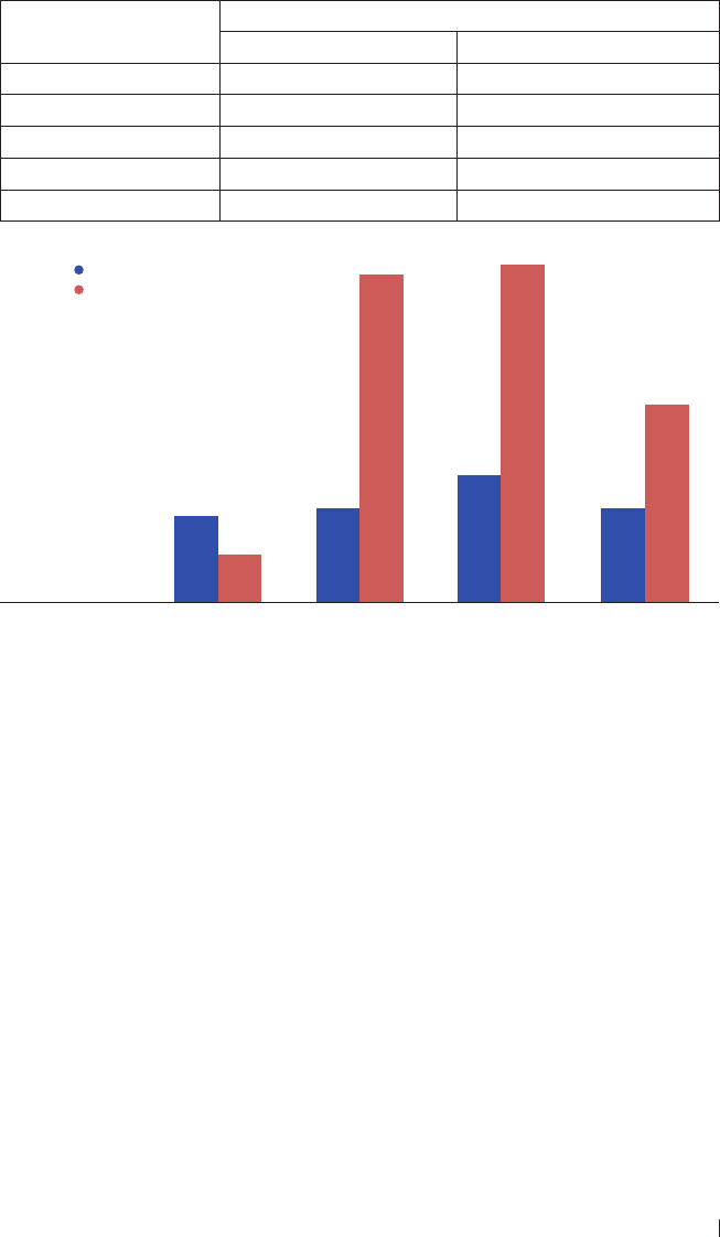
153
Acta Pharmaceutica Sciencia. Vol. 57 No. 2, 2019
Table 2. Number of paw licks (and percentage inhibition of licks) in rats administered with
formalin and pretreated with Combretum sordidum extract
Groups
Number of Licks
Early Phase (5 minutes) Late Phase (20-25minutes)
Control 107.5±17.07 (0%) 75.0±5.37 (0%)
CS 100mg/kg 90.3±15.09 (16.1%) 68.25±6.73 (9%)
CS 200mg/kg 91.3±8.08 (17.8%) 28.5±6.38 (62%)
CS 400mg/kg 81.8±3.89 (23.95%) 27.0±9.57 (64%)
Indomethacin 10mg/kg 88.5±1.03 (17.7%) 47.0±7.50 (37.3%)
Early phase (5 minutes)
Late phase (20-25 minutes)
% inhibition of licks
Figure 2. Number of paw licks (and percentage inhibition of licks) in rats administered with
formalin and pretreated with Combretum sordidum extract
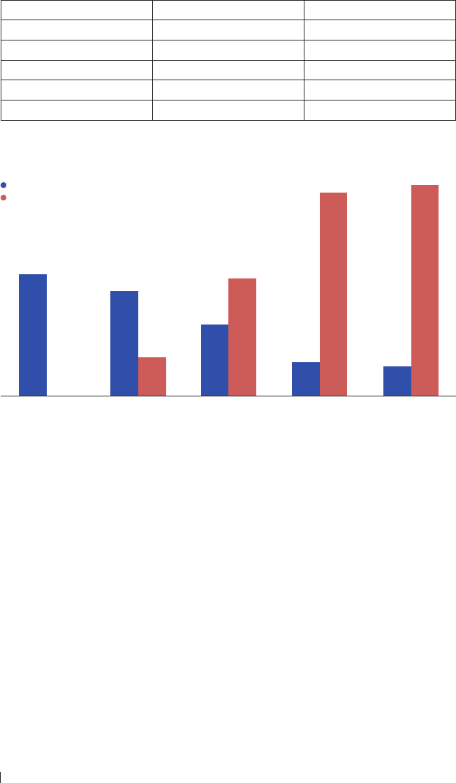
154
Acta Pharmaceutica Sciencia. Vol. 57 No. 2, 2019
Table 3. Abdominal writhing in mice intraperitonealy injected with acetic acid and pre-treated
with Combretum sordidum
Group Number of Writhes Inhibition (%)
Control 43.6±2.22 0
CS 100mg/kg 37.6±3.41 13.76
CS 200mg/kg 25.3±4.90 42.09
CS 400mg/kg 11.8±1.19 73.05
Indomethacin 10mg/kg 10.5±0.83 75.9
Figure 3. Abdominal writhing in mice intraperitonealy injected with acetic acid and pre-
treated with Combretum sordidum
Number of writhes
Inhibition

155
Acta Pharmaceutica Sciencia. Vol. 57 No. 2, 2019
Table 4. Effect of Methanol-water, ethyl acetate, and Hexane fractions on acetic acid- induced
abdominal writhing in mice
Group Number of writhes Inhibition (%)
Control 62.6 ± 1.25 0
Aspirin 15mg/kg 35.8 ± 4.31 42.8
MeOH 20mg/kg 39.0 ± 3.53 37.7
MeOH 50mg/kg 46.5 ± 2.97 25.7
EA 20mg/kg 35.4 ± 1.67 43.5
EA 50mg/kg 43.0 ± 4.10 31.3
Hex 20mg/kg 34.8 ± 4.14 44.4
Hex 50mg/kg 46.0 ± 3.65 26.5
Number of writhes
Inhibition
Figure 4. Effect of Methanol-water, ethyl acetate, and Hexane fractions on acetic acid-
induced abdominal writhing in mice
In this study, acetone extract of C. sordidum demonstrated anti-inamma-
tory activities by inhibition of paw oedema formation after exposure to egg
albumen and paw licking as a response to formalin-induced pain in rats. The
anti-inammatory response was comparable to that of indomethacin, a non-
steroidal anti-inammatory drug used for treatment of arthritis, gout, bursi-
tis.
18
Egg albumen-induced paw oedema is an animal model of inammation
used to detect anti-inammatory agents with activity in the acute phase of inf-
lammation.
19
This study demonstrated the anti-inammatory eect of C. sor-
didum was significantly established by 60 minutes post-injection of albumen.
This suggests anti-inammation with activity in acute inammatory phase.
Findings from the formalin-induced paw lick model also corroborated the an-
ti-inammatory eect of C. sordidum. It further established a more marked
anti-inammatory activity in the late phase of inammation, with significantly
more profound anti-inammatory action compared to the same indomethacin.
Formalin-induced paw lick model of inammation demonstrates the inter-
relation of inammation and nociception with distinct responses in the early

156
Acta Pharmaceutica Sciencia. Vol. 57 No. 2, 2019
and late phases of inammation but mediated via dierent inammatory
pathways.
20
The early phase is correlated with direct eect on peripheral no-
ciceptors with minimal inuence of prostaglandins. This stimulation in the
early phase culminates in centrally mediated pain accompanied by release of
substance P.
21,22
The late phase is characterized by release of mediators of inf-
lammation; histamine, prostaglandins, serotonin and bradykinin.
23,24
The an-
ti-inammatory activity of C. sordidum is suggested to be mediated via the pe-
ripheral and central mechanisms of inammatory pathways, and may involve
inhibition of cyclooxygenase enzymes which are active players in the model.
Centrally acting drugs such as opioids inhibit both phases equally but perip-
herally acting drugs such as cyclooxygenase inhibitors and corticosteroids only
inhibit the late phase.
25
The acetone extract of Combretum sordidum was also found to possess analge-
sic activities in this study with the significant inhibition of abdominal writhing
in the pretreated mice. Acetic acid- induced writhing in mice is a commonly
used model of analgesia in experimental animals for establishment of periphe-
rally acting analgesic drugs which may involve stimulation of local peritoneal
receptors.
26,27
The crude extract of C. sordidum showed a dose-dependent and
significant analgesic action when compared to the reference drugs, indomet-
hacin and aspirin. Acetic acid causes pain by liberating endogenous substances
that excite pain nerve endings.
28
Pain sensation in acetic acid–induced writhing method is elicited by trigge-
ring localized inammatory response resulting in release of free arachidonic
acid from tissue phospholipids via cyclooxygenase (COX) and prostaglandin
(PG) biosynthesis.
29,30
The acetic acid – induced writhing has been specifically
associated with increased level of PGE
2
and PGE
2
α in peritoneal uids as well
as lipooxygenase products.
31
The increase in prostaglandin levels within the
peritoneal cavity then enhances inammatory pain by increasing capillary
permeability. The acetic acid- induced writhing method is eective to evalu-
ate peripherally active analgesics. Agents which reduce the number of writhes
will render analgesic eect probably by inhibition of prostaglandin synthesis,
a peripheral mechanism of pain inhibition.
32
Therefore, the result of this study
shows that the extract possesses strong peripheral analgesic eect. Some of the
phytoconstituents of this plant such as avonoids, saponins, and tannins have
been demonstrated to have significant pharmacological activities.
33
Solvent partitioned fractions of C. sordidum extract (hexane, ethyl acetate and
methanol-water fractions) demonstrated potent analgesic eect in acetic acid-
induced writhing test in mice, with the hexane fraction showing the highest

157
Acta Pharmaceutica Sciencia. Vol. 57 No. 2, 2019
activity at 20mg/kg body weight. Further purification of the hexane fraction
yielded 7 fractions with fractions 6 and 7 showing marked inhibition of abdo-
minal writhing, almost 2-folds of that observed in aspirin.
Extract of C. sordidum at all doses showed anti-inammatory and analgesic
activities which are comparable to that exhibited by indomethacin or aspirin.
This study suggested that the anti-inammatory eect of C. sordidum may be
mediated via both peripheral and central mechanisms of inammation. The
solvent partitioned fractions showed hexane fraction had the most potent anal-
gesic activity and further purification yielded 7 fractions. Fractions 6 and 7
showed remarkable analgesic eect with almost 2-folds of that observed for
aspirin. The peripheral pathway for mediation of the analgesic eect of the
extract, solvent-partitioned fractions and column fractions of hexane fractions
was established also. This is a report of preliminary anti-inammatory and
analgesic activities of C. sordidum. Further studies are going on to purify, cha-
racterize and completely elucidate the structure of the bioactive compound in
this extract, particularly bioactive compounds responsible for the remarkable
analgesic activity of the fractions.
REFERENCES
1. Miguel, M. G. Antioxidant and Anti-Inammatory Activities of Essential Oils: A short revi-
ew. Molecules. 2010, 15, 9252-9287
2. Kusuma, I. W.; Kuspradini, H.; Arung, E. T.; Aryani, F.; Min, Y. H.; Kim, J. S.; Kim, Y. Bi-
ological Activity and Phytochemical Analysis of Three Indonesian Medicinal Plants, Murraya
koenigii, Syzygium polyanthum and Zingiber purpurea. J. Acupunct. Meridian Stud. 2011,
4, 75-79.
3. Iwalewa, E. O.; McGaw, L. J.; Naidoo, V.; Elo, J. N. Inammation: the foundation of dise-
ases and disorders. A review of phytomedicines of South African origin used to treat pain and
inammatory conditions. Afr. J. Biotechnol. 2007, 6, 2868-2885.
4. Plytycz, B.; Seijelid, R. From inammation to sickness: historical perspective. Arch. Immu-
nol. Ther. Exp. 2003, 51, 105-109.
5. Parham, C.; Chirica, M.; Timans, J.; Vaisberg, E.; Travis, M.; Cheung, J. Panz, S.; Zhang,
R.; Singh, K. P.; Vega, F.; To, W.; Wagner, J.; O’Farrell, A. M.; McClanahan, T.; Zurawski, S.;
Hannum, C.; Gorman, D.; Rennick, D. M.; Kastelein, R. A.; De Waal Malefyt, R.; Moore, K.
W. A. receptor for the heterodimeric cytokine IL-23 is composed of IL-12Rbeta1 and a novel
cytokine receptor subunit, IL-23R. J. Immunol. 2002, 168, 5699-5708.
6. Gao, H. M.; Hong, J. S. Why neurodegenerative diseases are progressive: uncontrolled
inammation drives disease progression. Trends Immunol. 2008, 29, 357-365.
7. Teresita Guardia, Alejandra Ester Rotelli, Ame´rico Osvaldo Juarez, Lilian Eugenia Pelzer
(2001) Anti-inammatory properties of plant avonoids. Eects of rutin, quercetin and hes-
peridin on adjuvant arthritis in rat. Farmaco. 2001, 56, 683–687.
8. Baricevic, D. S.; Sosa, R.; Della Loggia, A.; Tubaro, B.; Simonovska, A.; Krasna, A.; Zupan-

158
Acta Pharmaceutica Sciencia. Vol. 57 No. 2, 2019
cic A. Topical anti-inammatory activity of Salvia oicinalis L. leaves: the relevance of
ursolic acid. J. Ethnopharmacol. 2001, 75, 125–132.
9. Olaoluwa, O.; Ogunbor, F. Phytochemical screening, antimicrobial properties and essential
oil constituents of Combretum sordidum Exell. Int. J. Pharm. Sci. Res. 2015, 6, 1176-1180.
10. Beaudelaire K. P.; Luciano, B.; Remy, B. T.; Marius, M.; Télesphore, B. N.; Hee-Juhn, P.;
Kyung-Tae, L.; Leon, A. T. Polyhydroxyoleanane-type triterpenoids from Combretum molle
and their antiinammatory activity. Phytochem. Lett. 2008, 1, 183-187.
11. Teshome, T. D.; Ayana, A.; Sileshi, N.; Tadesse, W. Savanna land use and its eect on
woody plant species diversity in Borana, Southern Ethiopia. Sci. Technol. Arts Res. J. 2012,
1, 43-52.
12.Elo, J. N.; Katerere, D. R.; McGaw, L. J. The biological activity and chemistry of the sout-
hern African Combretaceae. J. Ethnopharmacol. 2008, 119, 686-699.
13. Aroke S. A.; Lyndy, J. M.; Esameldin, E. E.; Vinasan, N.; Jacobus, N. E. Polarity of extracts
and fractions of four Combretum (Combretaceae) species used to treat infections and gast-
rointestinal disorders in southern African traditional medicine has a major eect on dierent
relevant in vitro activities. J. Ethnopharmacol. 2014, 154, 339–350.
14. Magwenzi, R.; Nyakunu, C.; Mukanganyama, S. The Eect of Selected Combretum species
from Zimbabwe on the growth and drug eux systems of Mycobacterium aurum and Myco-
bacterium smegmatis. J. Micr. Biochem.Technol. 2014, S3, 003.
15. Moyo, R.; Chimponda, T.; Mukanganyama, S. Inhibition of hematopoietic prostaglandin
D2 synthase (H-PGDS) by an alkaloid extract from Combretum molle. BMC Complement.
Altern. Med. 2014, 14, 221.
16. Ojewole, J. A. Cardiovascular eects of mollic acid glucoside, a 1α-hydroxycycloartenoid
saponin extractive from Combretum molle R Br ex G Don (Combretaceae) leaf. Cardiovasc.
J. Afr. 2008, 19, 128–134.
17. Elo, J. N.; Famakin, J. O.; Katerere, D. R. P. Combretum woodii (Combretaceae) leaf ext-
racts have high activity against Gram-negative and Gram-positive bacteria. Afr. J. Biotechnol.
2005, 4, 1161-1166.
18. Rubin, B. R.; Burton, R.; Navarra, S.; Antigua, J.; Londoño, J.; Pryhuber, K. G.; Lund,
M.; Chen, E.; Najarian, D. K.; Petruschke, R. A.; Ozturk, Z. E.; Geba, G. P. Eicacy and safety
profile of treatment with etoricoxib 120 mg once daily compared with indomethacin 50 mg
three times daily in acute gout: A randomized controlled trial. Arthritis Rheum. 2004, 50,
598-606.
19. Amos, S. B.; Chindo, I.; Edmond, P. A.; Wambebe, C.; Gamaniel, K. Anti-inammatory
and anti-nociceptive eects of Ficus platyphylla in rats and mice. J. Herbs Spices Med.
Plants. 2002, 9, 47-53.
20. Oyebanji, B. O.; Saba, A. B.; Oridupa, O. A. Anti-nociceptive and anti-inammatory ac-
tivities of methanol extract of the leaves of Cajanus cajan. Niger. J. Physiol. Sci. 2014, 29,
183-188
21. Shah, B. N.; Seth, A. K.; Maheshwari, K. M. A review on medicinal plants as a source of
anti-inammatory agents. Res. J. Med. Plants. 2011, 5, 101-115.
22. Oridupa, O. A.; Saba, A. B. Relative anti-inammatory and analgesic activities of the who-

159
Acta Pharmaceutica Sciencia. Vol. 57 No. 2, 2019
le fruit, fruit bark, pulp and seed of Lagenaria breviora Roberty. J. Pharm. Toxicol. 2012,
7, 288 -297.
23. Zeashana, H.; Amresha, G.; Raoa, C. V.; Singh, S. Antinociceptive activity of Amaranthus
spinosus in experimental animals. J. Ethnopharmacol. 2009, 122, 492–496.
24. Alam, M. B.; Hossain, M. S.; Chowdhury, N. S.; Asadujjaman, M.; Zahan, R.; Islam, M. M.;
Mazumder, M. E. H.; Haque, M. E.; Islam, A. Antioxidant, anti-inammatory and antioyretic
activities of Trichosanthes dioica Roxb. Fruits. J. Pharm. Toxicol. 2011, 6, 440-453.
25. Amanlou, M.; Dadkhah, F.; Salehnia A.; Farsam H.; Dehpour A. R. An anti-inammatory
and anti-nociceptive eect of hydroalcoholic extract of saturejakhuzistanicajamzad extract. J.
Pharm. Sci. 2005, 8, 102-106.
26. Hasan, S. M. R; Hossain, M. M.; Akter, R.; Jamila, M.; Mazumder, M. E. H.; Alam, M.
A.; Faruque, A.; Rana, S.; Rahman, S. Analgesic activity of the dierent fractions of the aerial
parts of Commelina benghalensis Linn. Int. J. Pharmacol. 2010, 6, 63-67.
27. Ullah, M.; Showkat, N. U.; Ahmed, S. Evaluation of Momordica charantia L fruit extract
for analgesic and anti-inammatory activities using in vivo assay. Res. J. Med. Plant. 2011,
6, 236-244.
28. Duarte, I. D. G.; Nakamura, M.; Ferreira, S. H. Participation of the sympathetic system in
acetic acid-induced writhing in mice. Braz. J. Med. Biol. Res. 1988, 21, 341–343.
29. Ahmed, F.; Hossain, M. H.; Rahman, A. A.; Shahid, I. Z. Antinociceptive and sedative
eects of the bark of Cerberaodollam Gaertn. Orient. Pharm. Exp. Med. 2006, 6, 344-348.
30. Derardt, R.; Jougney, S.; Delevalccee, F.; Falhout, M. Release of prostaglandins E and F
in an algogenic reaction and its inhibition. Eur. J. Pharmacol. 1980, 51, 17-24.
31. Zakaria, Z. A.; Abdul Gani, Z. D. F. Antinociceptive, anti-inammatory, and antipyretic
properties of an aqueous extract of Dicranopteris linearis leaves in experimental animal mo-
dels. J. Nat. Med. 2008, 62, 179-187.
32. Ferdous, M.; Rouf, R.; Shilpi, J. A.; Uddin, S. J. Antinociceptive activity of the ethanolic
extract of Ficus racemosa Linn. (Moraceae). Orient. Pharm. Exp. Med. 2008, 8, 93-96.
33. Egharevba, H. O.; Odigwe, A. C.; Abdullahi, M. S.; Okwute, S. K.; Okogun, J. I: Phytoc-
hemical analysis and broad-spectrum antimicrobial activity of Cassia Occidentalis (whole
plant). N. Y. Sci. J. 2010, 3, 74-81.

160
Acta Pharmaceutica Sciencia. Vol. 57 No. 2, 2019

161
Acta Pharmaceutica Sciencia. Vol. 57 No. 2, 2019
Acta Pharm. Sci. Vol 57 No: 2. 2019
DOI: 10.23893/1307-2080.APS.05710
Combination of Cumulative Area Pre-
Processing and Partial Least Squares for
Handling Intensely Overlapping Binary and
Ternary Drug Systems
Yahya S Al-Degs
1*
, Amjad H. El-Sheikh
1
, Eman A. Abu Saaleek
1
, Reema A. Omeir
1
,
Musab A. Al-Ghodran
1
1 Chemistry Department, The Hashemite University, P. O. Box 150459 Zarqa 13115 Jordan
ABSTRACT
The analytical performance of cumulative area pre-processing (CAP), a recently
developed signal filtering method, along with multivariate calibration for quan-
tification of spectrally overlying drugs was outlined. The drug combinations con-
taining high level of paracetamol (PAR) in the presence of caeine (CAF), chlorp-
heniramine maleate (CHL), pseudoephedrine hydrochloride (PSE), phenylephrine
hydrochloride (PHE), and diphenhydramine hydrochloride (DPH). The tested
formulations were: PAR-CAF-PHE, PAR-CAF-PHE, and PAR-DRH. Based on net-
analyte signal calculations, the formulations exhibited intense overlapping 53-68%
for PAR-PSE-CHL, 55-95% for PAR-CAF-PHE, and 44% for PAR-DRH. For each
system, PLS latent variables were estimated using cross-validation technique and
more factors were needed for highly overlapping systems. PLS-CAP was found app-
licable for drugs quantification in all systems with excellent performance regard-
less the size of spectral overlapping and ratios of components in the formulation.
For PAR-PSE-CHL (ratio 300:30:2 mg/tablet), the ingredients were quantified by
CAP-PLS with satisfactorily recoveries (RSD, n = 3) 89.9% (3.1%), 104.6% (2.7%),
and 99.0% (1.5%) for PAR, PSE, and CHL, respectively. Both PLS and CAP-PLS
were demonstrated the same performance for binary system of modest overlapping
and no component available in low concentration.
Keywords: Signal Pre-processing; Spectral overlapping; Binary and Ternary Drug
Formulations; PLS calibration
*Corresponding Author: Dr. Yahya Al-Degs, e-mail: yahyaaldeqs@yahoo.com
Yahya S. Al-Degs ORCID Number: 0000-0002-9555-7594
Amjad H. El-Sheikh ORCID Number: 0000-0001-8321-3236
Eman A. Abu Saaleek ORCID Number: 0000-0003-3163-2919
Reema A. Omeir ORCID Number: 0000-0002-3508-5927
Musab A. Al-Ghodran ORCID Number: 0000-0002-4766-0730
(Received 29 December 2018, accepted 24 January 2019)

162
Acta Pharmaceutica Sciencia. Vol. 57 No. 2, 2019
INTRODUCTION
Commercial drug formulations often contain more than one activates pharma-
ceutical ingredients (APIs) that present in variable levels to achieve the best
pharmacological performance
1
. The spectral overlapping between the active
ingredients is often moderate but intense overlapping is also possible. In ad-
dition to spectral overlapping and nonlinearity in the system, drug producti-
on stages like crystallization, drying, solid dosage form, added excipients, and
tableting at dierent conditions can aect the spectral behavior of APIs
1,2
. Ac-
cordingly, accurate analytical methods are always needed in this regard
1
. The
development and formulation of the pharmaceuticals brought a revolution in
human health. These pharmaceuticals would help their intent only if they are
free from impurities and are administered in an appropriate quantity. Hen-
ce, there are many challenges and it can be reduced by eective use of exci-
pients, which permits formulators to overcome these challenges. It becomes
necessary to develop new analytical methods because sometimes the dosage
form contains other substances which potentially interfere in the assay and, if
not corrected, may impart a systematic error to the assay
1
. Multi-component
formulations have gained a lot of attention nowadays due to greater patient
acceptability, increased potency, multiple action, quick relief and fewer side
eects
1,2
. Market is ooded with combination of drugs in various dosage form.
One of such combination is paracetamol (PAR) with other drugs including caf-
feine (CAF), diphenhydramine hydrochloride (DPH), chlorpheniramine ma-
leate (CHL), pseudoephedrine hydrochloride (PSE), phenylephrine hydrochlo-
ride (PHE). For example, PAR and DPH are co-formulated in pharmaceutical
product for temporary relief of pain when associated with sleeping diiculty
2-7
.
The combination PAR-CHL-PSE has been recently introduced in the market to
treat the symptoms of most ues
5-7
. The combination PAR-CAF-PHE was also
formulated to relief from major cold and u symptoms and applied at day time
as it doesn’t cause drowsiness.
Various chemical and instrumental methods were developed to make drugs
serve their purpose at regular intervals which are involved in the estimation of
drugs. These pharmaceuticals may develop impurities at various steps of the-
ir development, transportation and storage, which makes the pharmaceutical
risky to be administered. Thus, they must be identified and quantified. Hence,
analytical instrumentation and methods play an important role. Thus, the re-
view highlights a variety of analytical, chromatographic and instrumental met-
hod developed such as; High Performance Liquid Chromatography (HPLC),
High Performance Thin Layer Chromatography (HPTLC), and Gas Chroma-

163
Acta Pharmaceutica Sciencia. Vol. 57 No. 2, 2019
tography (GC) have wide application in assuring the quality and quantity of
pharmaceutical products and these instrumental methods are simple, precise,
rapid, and reproducible and have been applied in the analysis of pharmaceuti-
cals for assessing the quality of the drugs
8-10
.
Recently, multivariate calibration methods have been intensively employed for
analyzing pharmaceutical formulations and detection of released drugs in uri-
ne and plasma
5-7
. Particularly speaking, PAR and CAF have been quantified in
drug formulations using first-order multivariate calibration methods with high
accuracy and without implementing any chromatographic procedure
6,7
. Com-
pared to liquid chromatography, assaying drug formulations by multivariate
calibration required less solvent consumption and avoids using tedious chro-
matographic instruments
2,3
. First order multivariate calibration methods inc-
ludes multilinear regression (MLR), principal component regression (PCR),
partial least-squares (PLS-1), and many others
11
. In a recent study, both PAR
and PHE were quantified in four-drug formulations using PLS1 calibration
5,6
.
PHE was quantified in the presence of PAR in tablet form with excellent re-
covery
7
. In another study, novel application of PCR and PLS was developed
for determination of PAR, CAF, and CHL in real formulations containing ot-
her medicines
6,7
. In the drug formulation, the recovery and RSD values were
99.91% (1.69%), 100.15%(1.35%) and 100.49%(1.66%) for PAR, CAF and CHL,
respectively. In the second method PLS was applied to quantify four medicines
in real formulation with high recoveries and RSD: 99.80% (1.67%), 100.19%
(1.42%) and 100.45% (1.86%) for PAR, CAF and CHL, respectively
7
. There is
no published literature dealing with simultaneous quantification of PAR and
DPH in real formulations. Spectral overlapping between drugs was reported to
have a negative inuence on the performance of multivariate calibration met-
hods
11
. Form the earlier studies, it is concluded that multivariate calibration is
applicable for medicines determination in dierent commercial formulations
including tablet and syrups.
Usually, the pre-processing methods are divided into three categories: a) filte-
ring and de-noising, generally associated with the improvement of the signal-
to-noise ratio. These methods are applied to the raw data before the construc-
tion of the multivariate model and reduce the inuence of the random variance
without altering that useful, b) spectral normalization and dierentiation,
such as the spectral derivation and the application of the Fourier transform.
These procedures are also applied before the construction of the model, and
c) selection of the variables and reduction of dimensionality of the data. These
methods are applied by multivariate modeling and therefore involve both the

164
Acta Pharmaceutica Sciencia. Vol. 57 No. 2, 2019
matrix X (independent variables) and the matrix Y (dependent variables). The
most used method is orthogonal signal correction. There are a number of ex-
cellent review articles providing guidance for application of the pre-processing
techniques to the analytical signals
12-14
. In some pharmaceutical formulations
one or more components are present in very low concentrations, so as to be
hidden by the components more concentrated or by the instrumental noise.
This increases the diiculty of building a mathematical model able to predict in
a satisfactory manner all the species present in the mixture. In these cases, the
data pre-treatment should provide to amplify the analytical information due to
the components at lower concentration and at the same time to minimize the
spectral noise that can interfere with their determination.
In the current work, the application of newly proposed cumulative area pre-
processing (CAP) will be evaluated for prior to PLS calibration for quantifica-
tion of binary and ternary drugs systems. CAP was developed to amplify the
spectral signals. CAP-PLS will be tested on the quantitative analysis of one
binary and ternary pharmaceutical formulation. The common diiculty in
analyzing these matrices was the presence of some components in much lower
amount than other components. The binary formulation consisted of para-
cetamol-diphenhydramine hydrochloride (ratio 20:1), paracetamol-caeine-
phenylephrine hydrochloride (ratio 100:5:1 mg/tablet), and paracetamol-pse-
udoephedrine hydrochloride-chlorpheniramine maleate (ratio 150:15:1 mg/
tablet). The analytical performance of CAP-PLS and PLS for drugs quantifica-
tion will critically discussed. More attention will be paid on the inuence spect-
ral overlapping among drugs on their quantification by CAP-PLS. Inuence of
pH and spectral range on spectral analysis will be also considered.
METHODOLOGY
Drugs and marketed formulations
The pharmaceuticals paracetamol (PAR), caeine (CAF), diphenhydramine
hydrochloride (DPH), phenylephrine hydrochloride (PHE), pseudoephedrine
hydrochloride (PSE), and chlorpheniramine maleate (CHL) were kindly do-
nated from Dar Al-Dawaa company (Dar Al-Dawaa, Naour, Amman, Jordan).
Distilled water was used for preparation of standard solutions and dissoluti-
on of commercial tablets. The marketed drugs were Panadol cold and u®
(Active ingredients: PAR-PSE-CHL 500:30:2 mg/tablet; excipients Silicon di-
oxide, stearic acid, sodium benzoate, povidone, starch, maize starch, and talc,
GlaxoSmithKline Dungarvan Ltd., Co. Waterford, Ireland), Panadol cold and
u Day® (Active ingredients: PAR-CAF-PHE 500:25:5 mg/tablet; excipients
pre-gelatinised starch, maize starch, povidone, potassium sorbate, talc, stearic
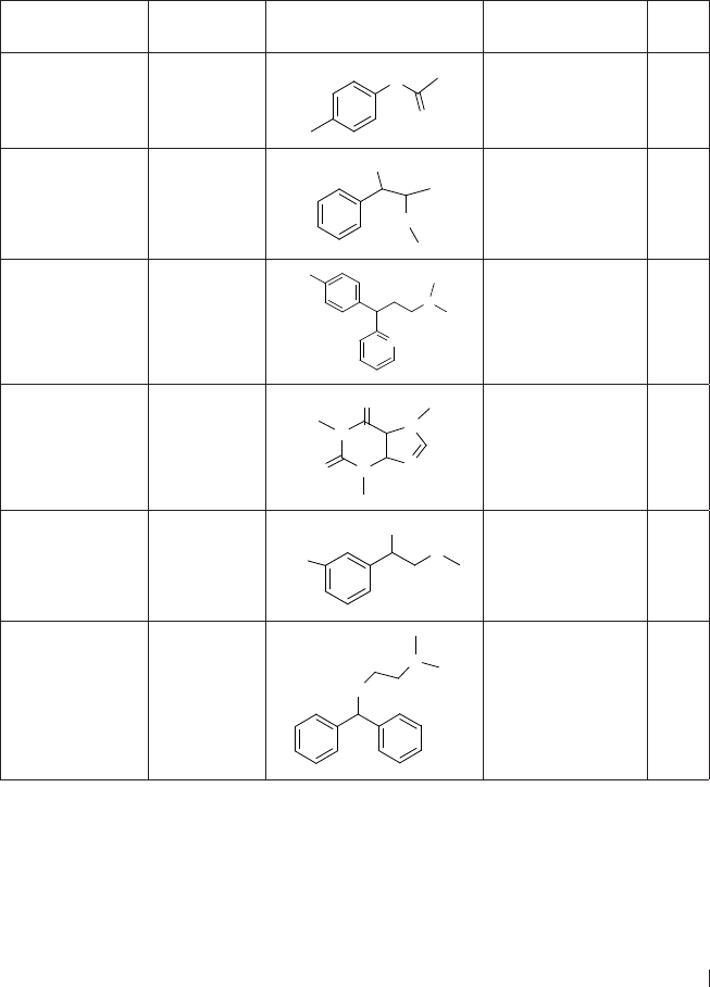
165
Acta Pharmaceutica Sciencia. Vol. 57 No. 2, 2019
acid, microcrystalline cellulose, sodium lauryl sulphate, sunset yellow, Gla-
xoSmithKline Dungarvan Ltd., Co. Waterford, Ireland), and Panadol night
®
(PAR-DPH 500:25 mg/tablet; excipients maize starch, pregelatinised starch,
potassium sorbate, povidone, talc, and stearic acid, GlaxoSmithKline Dungar-
van Ltd., Co. Waterford, Ireland) All other reagents were of the highest purity
commercially available. Table 1 summaries some chemical parameters of the
undertaken pharmaceuticals.
Table 1. Chemical properties of studied pharmaceuticals.
Name/short
name
Formula Structural formula Medical action pK
a
Paracetamol
PAR
C
8
H
9
O
2
N
H
O
OH
Treat mild to
moderate pains
9.5
Pseudoephedrine
hydrochloride
PSE
C
10
H
15
ON
NH
OH
Shrink blood
vessels in the nasal
passage (prevent
stuffy nose)
10.3
Chlorpheniramine
Maleate
CHL
C
15
H
19
N
2
Cl
N
N
Cl
Relieve symptoms
of allergy
9.1
Caffeine
CAF
C
8
H
10
N
4
O
2
N
N
N
N
O
O
Acts as central
nervous system
stimulant
14.0
Phenylephrine
hydrochloride
PHE
C
9
H
14
ClNO
2
OH
N
H
OH
H Cl
Sympathomimetic
(descongestants)
9.07
Diphenhydramine
Hydrochloride
DPH
C
17
H
22
ClNO
O
N
H Cl
Blocking the effects
of histamine and
causes drowsiness.
9.13

166
Acta Pharmaceutica Sciencia. Vol. 57 No. 2, 2019
Standard solutions, calibration/validation mixtures
Stock solutions were separately prepared by dissolving in ethanol nearly 25.0
(±0.0001 g) mg of each drug in 100 mL volumetric asks. A first set of 9 binary
calibration samples was built by combining five levels of PAR and DPH within
the range 1.0–12.0 mg/L.A second calibration set of nine ternary mixture solu-
tions was prepared for PAR-CAF-PHE and PAR-PSE-CHL systems. For PAR-
CAF-PHE, drug ranges were 2.0-15.0 mg/L
for PAR, 2.0-16.0 mg/L for CAF
and 1.0-8.0mg/L for PHE. For PAR-PSE-CHL, drug ranges were1.0-16.0 mg/L
for PAR, 1.0-8.0 mg/L for PSE, and 1.0-12.0 mg/L for CHL. Calibration set of
the binary mixtures was created by following a full experimental design while
calibration sets of binary systems selected according to Brereton`s rule using
three dierent concentration levels. To validate the PLS and CAP-PLS models,
three independent external validation sets consisting of 7 solutions for binary
and ternary systems were prepared. Marketed formulations were assayed by
weighing four tablets for binary system and five tablets for ternary systems
and grinding the tablets to a fine powder. The powder was suspended in water
and diluted to a final volume of 100 ml. The suspension was sonicated for 10
min, filtered through a 0.45μm filter, and properly diluted in preparation for
spectral analysis.
Partial least squares and net-analyte signal calculations
A double beam UV-visible spectrophotometer (Thermo scientific. Genesys
10S UV-VIS (USA)) with quartz cuvette cell of 1.0 cm path length was emplo-
yed. The spectral bandwidth was 1.0 nm with fast wavelength-scanning speed.
Scans were carried out in the range of 200–300 nm at (1.0 nm step, 101 points/
spectrum). The spectral data obtained for calibration mixtures are placed in
matrix A (size 101×9) and spectral data of validation set are placed matrix B
(size 101×7). On the other hand, concentration data are placed matrix C (size
9×3 for ternary systems and size 9×2 for binary system). Partial least squa-
res PLS1 is an eicient tool for developing a quantitative relationship between
several predictor variables A (spectral data) and a property of interest c (the
independent variable or drugs content) as shown below
11,15
:
c=Ab (1)
Where c contains the concentrations of calibrated drug in calibration samples
arranged in a vector and b is the calibration sensitivity which is necessary for
estimating drugs content in the adsorption solutions
11,16-18
:
b=W
t
(PW
t
)
-1
q (2)

167
Acta Pharmaceutica Sciencia. Vol. 57 No. 2, 2019
Where W is the weights matrix for A, P is the loadings matrix of A, q is the
loadings vector for c, t donates transpose operation, and -1 stands for inverse
operation. Once b is estimated, prediction of drug concentrations (c
un
) from
the unknown spectrum a is carried out as following
11,16-18
:
c
un
= ab (3)
Optimum number of PLS-latent variables were estimated using cross-validation
method
15
. The predicted concentrations were compared with the known con-
centrations of the compounds in each calibration sample. The prediction error
sum of square (PRESS) and the relative error of prediction (REP %) were cal-
culated for drugs in calibration and validation samples as following
17,19
:
Partial least squares and net-analyte signal calculations
A double beam UV-visible spectrophotometer (Thermo scientific. Genesys 10S UV-VIS (USA)) with
quartz cuvette cell of 1.0 cm path length was employed. The spectral bandwidth was 1.0 nm with fast
wavelength-scanning speed. Scans were carried out in the range of 200–300 nm at (1.0 nm step, 101
points/spectrum). The spectral data obtained for calibration mixtures are placed in matrix A (size
101×9) and spectral data of validation set are placed matrix B (size 101×7). On the other hand,
concentration data are placed matrix C (size 9×3 for ternary systems and size 9×2 for binary system).
Partial least squares PLS1 is an efficient tool for developing a quantitative relationship between
several predictor variables A (spectral data) and a property of interest c (the independent variable or
drugs content) as shown below
11,15
:
c=Ab (1)
Where c contains the concentrations of calibrated drug in calibration samples arranged in a vector and
b is the calibration sensitivity which is necessary for estimating drugs content in the adsorption
solutions
11,16-18
:
b=W
t
(PW
t
)
-1
q (2)
Where Wis the weights matrix for A, P is the loadings matrix of A, q is the loadings vector for c, t
donates transpose operation, and -1 stands for inverse operation. Once b is estimated, prediction of
drug concentrations (c
un
) from the unknown spectrum a is carried out as following
11,16-18
:
c
un
= ab (3)
Optimum number of PLS-latent variables were estimated using cross-validation method
15
. The
predicted concentrations were compared with the known concentrations of the compounds in each
calibration sample. The prediction error sum of square (PRESS) and the relative error of prediction
(REP %) were calculated for drugs in calibration and validation samples as following
17,19
:
=
−=
n
i
prediactuali
CCPRESS
1
2
,,
)(
(4)
(4)
%REP = 100×
%REP = 100×
2/1
1
2
,
2
,
1
,
)(
)(
−
=
=
m
i
acti
acti
n
i
predi
C
CC
(5)
Where C
actual
, C
prd
, and n, are actual concentration (mg/L), predicted concentration obtained by PLS
(mg/L), and number of training samples (solutions), respectively. A unifying framework for
calibration and prediction in multivariate calibration is shown based on the concept of the Net Analyte
Signal (NAS). NAS is the part of the measured spectrum that the calculated model uses for prediction.
The main equation that is needed to estimated figures of merit is
15,20,21
:
s
k
* = [I-S
-k
S
-k
+
]s
k
(6)
Where Sis the matrix of sensitivities collected for all other solutes, s
k
is the sensitivity vector of the
analyte, and s
k
* is the spectrum of pure analytek measured at unite concentration (sensitivity factor).
Sensitivity (SEN) value of analytek is the norm (║║) of the net sensitivity vector, it was defined as the
amount of net signal that, in prediction, corresponds to a concentration equal to unity
21
:
SEN = ║s
k
*║ (7)
Selectivity (SEL) can be defined as the part of the measured signal unique to the analyte of interest.
Based on NAS theory, the selectivity measures the extent of spectral overlapping. SEL can be
expressed by the ratio between the norm of the NAS vector and the norm of the spectra. SEL values
are extended from zero (high overlap with other interferences) to unity (no overlap with other sample
components). High values of SEN are an indication of high method accuracy for that analyte
15,20,21
:
SEL = ║s
k
*║/║s
k
║ (8)
The limit of detection (LOD) obtained assumes that the prediction uncertainties are approximately
constant. LOD gives the minimum detectable amount of k solute. A reasonable estimation to
determine the LOD can be estimated as
21
:
LOD = 3║ε║/║s
k
*║ (9)
Where ║ε║ is a measurement of the instrumental noise, and s* was defined above. Then the norms of
blank readings (║NAS
blank
║) are estimated and ║ε║ is taken as the standard deviation of estimated
norms
21
.The limit of quantification (LOQ) is the minimum quantifiable amount of the solute, is
estimated as
15,21
:
(5)
Where C
actual
, C
prd
, and n, are actual concentration (mg/L), predicted concent-
ration obtained by PLS (mg/L), and number of training samples (solutions),
respectively. A unifying framework for calibration and prediction in multi-
variate calibration is shown based on the concept of the Net Analyte Signal
(NAS). NAS is the part of the measured spectrum that the calculated model
uses for prediction. The main equation that is needed to estimated figures of
merit is
15,20,21
:
s
k
* = [I-S
-k
S
-k
+
]s
k
(6)
Where Sis the matrix of sensitivities collected for all other solutes, s
k
is the sen-
sitivity vector of the analyte, and s
k
* is the spectrum of pure analytek measured
at unite concentration (sensitivity factor). Sensitivity (SEN) value of analytek
is the norm (║║) of the net sensitivity vector, it was defined as the amount of
net signal that, in prediction, corresponds to a concentration equal to unity
21
:
SEN = ║s
k
*║ (7)
Selectivity (SEL) can be defined as the part of the measured signal unique to
the analyte of interest. Based on NAS theory, the selectivity measures the ex-
tent of spectral overlapping. SEL can be expressed by the ratio between the
norm of the NAS vector and the norm of the spectra. SEL values are extended

168
Acta Pharmaceutica Sciencia. Vol. 57 No. 2, 2019
from zero (high overlap with other interferences) to unity (no overlap with ot-
her sample components). High values of SEN are an indication of high method
accuracy for that analyte
15,20,21
:
SEL = ║s
k
*║/║s
k
║ (8)
The limit of detection (LOD) obtained assumes that the prediction uncertainti-
es are approximately constant. LOD gives the minimum detectable amount of
k solute. A reasonable estimation to determine the LOD can be estimated as
21
:
LOD = 3║ε║/║s
k
*║ (9)
Where ║ε║ is a measurement of the instrumental noise, and s* was defined
above. Then the norms of blank readings (║NAS
blank
║) are estimated and ║ε║
is taken as the standard deviation of estimated norms
21
.The limit of quantifi-
cation (LOQ) is the minimum quantifiable amount of the solute, is estimated
as
15,21
:
LOQ = 10║ε║/║s
k
*║ (10)
All calculations were carried out using special matlab-codes under MATLAB
®
.
Cumulative area pre-processing of spectral data prior to
PLS calibration
Data treatment by CAP transforms the original UV spectrum to a new curve
using the area underlying the same spectrum. This elaboration is based on two
mathematical steps. In the first step, the area a
λi
binning of the two consecutive
wavelengths (λ
i
and λ
i+1
) is calculated as
7
:
LOQ = 10║ε║/║s
k
*║ (10)
All calculations were carried out using special matlab-codes under MATLAB
®
.
Cumulative area pre-processing of spectral data prior to PLS calibration
Data treatment by CAP transforms the original UV spectrum to a new curve using the area underlying
the same spectrum. This elaboration is based on two mathematical steps. In the first step, the area a
λi
binning of the two consecutive wavelengths (λ
i
and λ
i+1
) is calculated as
7
:
2
1+
+
=
ii
i
abab
a
(11)
Where ab is the absorbance value at a single wavelength. This calculation is repeated for all the
wavelengths in the spectrum. In the second step, the cumulative sum of the single areas is computed,
in such a way as the cumulative area at each wavelength A
λi
resulted from the sums of all areas a
i
of
the preceding wavelengths. For a spectral region between λ
m
and λ
n
(λ
m
<λ
n
), the cumulative area A
λm,n
is estimated as
7
:
=
=
n
mi
inm
aA
,
(12)
Treatment of the original UV spectra using CAP (Eqs 11 and 12) offers many advantages including
amplification of the analytical signals for all solutes and possible quantification
RESULTS AND DISCUSSION
Influence of solution pH on spectral behavior of pharmaceuticals
Influence of solution pH on spectral behavior was studied for all drugs at pH 3.0, 7.0, and 10.0 to
cover all possible conditions. Among the drugs, identical spectral shapes over the range (230-280 nm)
were observed for CHL and PSE and hence there was no need to present their profiles. On the other
hand, the UV-absorption behavior of PAR (which present in all systems), CAF, PHE, and DPH were
notably changed with solution pH as depicted in Fig 1.
(11)
Where ab is the absorbance value at a single wavelength. This calculation is
repeated for all the wavelengths in the spectrum. In the second step, the cumu-
lative sum of the single areas is computed, in such a way as the cumulative area
at each wavelength A
λi
resulted from the sums of all areas a
i
of the preceding
wavelengths. For a spectral region between λ
m
and λ
n
(λ
m
<λ
n
), the cumulative
area A
λm,n
is estimated as
7
:
LOQ = 10║ε║/║s
k
*║ (10)
All calculations were carried out using special matlab-codes under MATLAB
®
.
Cumulative area pre-processing of spectral data prior to PLS calibration
Data treatment by CAP transforms the original UV spectrum to a new curve using the area underlying
the same spectrum. This elaboration is based on two mathematical steps. In the first step, the area a
λi
binning of the two consecutive wavelengths (λ
i
and λ
i+1
) is calculated as
7
:
2
1+
+
=
ii
i
abab
a
(11)
Where ab is the absorbance value at a single wavelength. This calculation is repeated for all the
wavelengths in the spectrum. In the second step, the cumulative sum of the single areas is computed,
in such a way as the cumulative area at each wavelength A
λi
resulted from the sums of all areas a
i
of
the preceding wavelengths. For a spectral region between λ
m
and λ
n
(λ
m
<λ
n
), the cumulative area A
λm,n
is estimated as
7
:
=
=
n
mi
inm
aA
,
(12)
Treatment of the original UV spectra using CAP (Eqs 11 and 12) offers many advantages including
amplification of the analytical signals for all solutes and possible quantification
RESULTS AND DISCUSSION
Influence of solution pH on spectral behavior of pharmaceuticals
Influence of solution pH on spectral behavior was studied for all drugs at pH 3.0, 7.0, and 10.0 to
cover all possible conditions. Among the drugs, identical spectral shapes over the range (230-280 nm)
were observed for CHL and PSE and hence there was no need to present their profiles. On the other
hand, the UV-absorption behavior of PAR (which present in all systems), CAF, PHE, and DPH were
notably changed with solution pH as depicted in Fig 1.
(12)

169
Acta Pharmaceutica Sciencia. Vol. 57 No. 2, 2019
Treatment of the original UV spectra using CAP (Eqs 11 and 12) oers many
advantages including amplification of the analytical signals for all solutes and
possible quantification
RESULTS AND DISCUSSION
Inuence of solution pH on spectral behavior of pharmaceuticals
Inuence of solution pH on spectral behavior was studied for all drugs at pH
3.0, 7.0, and 10.0 to cover all possible conditions. Among the drugs, identical
spectral shapes over the range (230-280 nm) were observed for CHL and PSE
and hence there was no need to present their profiles. On the other hand, the
UV-absorption behavior of PAR (which present in all systems), CAF, PHE, and
DPH were notably changed with solution pH as depicted in Fig 1.
Figure 1. Influence of pH on spectral behavior of tested drugs (7.0 mg/Leach drug)
As shown in Fig 1, large variation was noted in the spectra of CAF at the studied pH and over the
range (200-240 nm) and slight variations were observed over the rest of the domain. In the same time,
the distinct wavelength positioned at 273 nm was not affected with solution pH. More variations were
observed in the PAR spectra, the intensity was notably increased at pH over the range 200-220 nm
while the main peak (243 nm) was shifted to 250 nm at pH 10.0. The most drastic changes were
reported for PHE as shown in Fig 1. It was highly possible that PHE underwent chemical reaction at
the basic medium and this was deduced from the developed spectrum which has two wavelengths at
208 and 237 nm. To avoid PHE side-reaction, it was practical to run UV measurements at pH 7.0. As
can be noted from the earlier discussion, the optimum pH for spectral analysis is 7.0 which maintain
stable structure and high UV absorption for all drugs. For DPH, there was a significant light
absorption with pH over the range (200-230 nm), while, pH has no effect on the spectrum over the rest
of the domain. pH 7.0 seems to be a good choice to run spectral measurement for this drug. In fact,
protonation, hydrolysis, and internal-arrangement of drug molecules are highly possible at acidic or
0,0
0,5
1,0
1,5
200 220 240 260 280 300
Absorbance
Wavelength/nm
CAF
pH 3
pH 7
pH 10
0,0
0,5
1,0
1,5
200 220 240 260 280 300
Absorbance
Wavelength/nm
PAR
pH 3
pH 7
pH 10
0
0,1
0,2
0,3
0,4
0,5
0,6
0,7
200 220 240 260 280 300
Absorbance
Wavelength/nm
PHE
pH 3
pH 7
pH 10
0,0
0,2
0,4
0,6
0,8
1,0
1,2
200 220 240 260 280 300
Absorbance
Wavelength/nm
DPH
pH 3
pH 7
pH 10
Figure 1. Inuence of pH on spectral behavior of tested drugs (7.0 mg/Leach drug)
As shown in Fig 1, large variation was noted in the spectra of CAF at the studied
pH and over the range (200-240 nm) and slight variations were observed over the
rest of the domain. In the same time, the distinct wavelength positioned at 273
nm was not aected with solution pH. More variations were observed in the PAR
spectra, the intensity was notably increased at pH over the range 200-220 nm
while the main peak (243 nm) was shifted to 250 nm at pH 10.0. The most drastic

170
Acta Pharmaceutica Sciencia. Vol. 57 No. 2, 2019
changes were reported for PHE as shown in Fig 1. It was highly possible that PHE
underwent chemical reaction at the basic medium and this was deduced from the
developed spectrum which has two wavelengths at 208 and 237 nm. To avoid PHE
side-reaction, it was practical to run UV measurements at pH 7.0. As can be noted
from the earlier discussion, the optimum pH for spectral analysis is 7.0 which ma-
intain stable structure and high UV absorption for all drugs. For DPH, there was a
significant light absorption with pH over the range (200-230 nm), while, pH has
no eect on the spectrum over the rest of the domain. pH 7.0 seems to be a good
choice to run spectral measurement for this drug. In fact, protonation, hydrolysis,
and internal-arrangement of drug molecules are highly possible at acidic or basic
solutions and this aects their spectral properties. Accordingly, solution pH sho-
uld be adjusted to get unique spectral characteristics of the measured drug.
Spectral behavior of pharmaceuticals and spectral overlapping
In fact, intense spectral overlapping among pharmaceuticals would retard their qu-
antification in solution and in real formulation
11,15
. Accordingly, the spectral behavior
of pharmaceuticals and extent of overlapping were evaluated before running mul-
tivariate calibration. The UV spectra of the studied systems are presented in Fig. 2
basic solutions and this affects their spectral properties. Accordingly, solution pH should be adjusted
to get unique spectral characteristics of the measured drug.
Spectral behavior of pharmaceuticals and spectral overlapping
In fact, intense spectral overlapping among pharmaceuticals would retard their quantification in
solution and in real formulation
11,15
. Accordingly, the spectral behavior of pharmaceuticals and extent
of overlapping were evaluated before running multivariate calibration. The UV spectra of the studied
systems are presented in Fig. 2
Figure 2. UV Spectra of the binary and ternary drug systems. Top PAR–DPH (6.0 mg/L and pH 7.0),
Middle CAF–PAR–PHE (7.0 mg/L and pH 7.0) Bottom PAR–PSE–CHL (5.0 mg/L and pH 7.0)
As indicated in Fig. 2, all drugs exhibited strong light absorption over the range (200 300 nm) in
addition to intense overlapping among the signals. For PAR-DPH, a large spectral overlap was
observed between both compounds and particularly over the range 220-280 nm. In addition to intense
overlapping, PAR has better UV absorption compared to DPH and would make their quantification by
0,0
0,2
0,4
0,6
0,8
1,0
1,2
1,4
200 220 240 260 280 300
Absorbance
Wavelength/nm
Paracetamol (PAR)
Diphenhydramine
hydrochloride (DPH)
0,00
0,50
1,00
1,50
200 220 240 260 280 300
Absorbance
Wavelength/nm
Caffeine (CAF)
Paracetamol (PAR)
Phenylephrine Hydrochloride
(PHE)
0,00
0,10
0,20
0,30
0,40
0,50
0,60
0,70
200 220 240 260 280 300
Absorbance
Wavenumber/nm
Paracetamol (PAR)
Pseudoephedrine hydrochloride (PSE)
Chlorpheniramine Maleate (CHL)
Figure 2. UV Spectra of the binary and ternary drug systems. Top PAR–DPH (6.0 mg/L and
pH 7.0), Middle CAF–PAR–PHE (7.0 mg/L and pH 7.0) Bottom PAR–PSE–CHL (5.0 mg/L and
pH 7.0)

171
Acta Pharmaceutica Sciencia. Vol. 57 No. 2, 2019
As indicated in Fig. 2, all drugs exhibited strong light absorption over the range
(200 300 nm) in addition to intense overlapping among the signals. For PAR-
DPH, a large spectral overlap was observed between both compounds and par-
ticularly over the range 220-280 nm. In addition to intense overlapping, PAR
has better UV absorption compared to DPH and would make their quantificati-
on by simple spectrometry (especially DPH) a hard task. The intense absorpti-
on of PAR at 243 nm was mainly attributed to the n-π*. One more point on this
PAR-DPH is the presence of DPH in much lower amount compared to PAR in
real formulation and this added more analytical obstacles on their accurate qu-
antification. For the ternary system CAF–PAR–PHE, the solutes were actively
absorbing over the region this was attributed to their chemical structure which
contains many active functional groups. The spectra of the drugs indicated the
following distinct absorption wavelengths at 243, 273, and (215 and 272 nm)
for PAR, CAF and PHE, respectively. The reported absorption wavelengths
were mainly attributed to n→π* and π→π* electronic transitions in the mole-
cules. In fact, the longer maximum wavelength of absorption observed for CAF
(273 nm) was attributed to the conjugated system as indicated from the chemi-
cal structures of the drugs (Table 1). Generally, PHE showed weaker absorption
compared to other drugs and absorb near the lower end of the spectral range
(220 nm). The spectral overlap between drugs is high over the entire spectral
range. Within the spectral regions 250-270, an intense overlapping between
PAR and CAF was observed and this would aect their quantification in real
formulations. Moreover, PHE absorbs over the entire range. In this ternary
system, PHE should be added in much lower levels and this negatively reected
on its quantification in real formulations. For PAR–PSE–CHL, the solutes were
active in UV region and this is attributed to their chemical structure which con-
tains aromatic parts substituted with functional groups (Table 1). The spectra
of drugs indicated the following special absorption wavelengths 210, 242, and
265 nm for PSE, PAR and CHL, respectively. The observed bands were mainly
attributed to n→π* and π→π*electronic transitions. Generally, PSE has a poor
UV absorption compared to two drugs and absorb near the extreme of the app-
lied spectral domain. In fact, the spectral overlap between drugs is high over
the entire spectral range. Within the spectral regions 220-240 and 260-280
nm, an intense overlapping between PAR and CHL was observed (Fig. 1). Mo-
reover, PSE absorbs over the entire range. Practically, PAR and PSE are added
in much higher levels compared to CHL and this would make quantification
of the later drug a hard-analytical task. For each system, the extent of spectral
overlapping between drugs was estimated using net-analyte signal calculations
(Eq. 8) and the results are provided in Table 2.

172
Acta Pharmaceutica Sciencia. Vol. 57 No. 2, 2019
Table 2. Extent of spectral overlapping in the drug systems using NAS calculations
a
Total spectral overlapping with other drugs%
PAR–PSE–CHL
PAR 53
PSE 55
CHL 68
CAF–PAR–PHE
CAF 68
PAR 85
PHE 79
PAR–DPH
PAR 38
DPH 62
a
For PAR–PSE–CHL, NAS calculations were carried out at 5.0 mg/L, pH 7.0, and spectral
range 200-300 nm. For CAF–PAR–PHE, calculations were carried out at 7.0 mg/L, pH 7.0 and
200-300 nm. For PAR–DPH, 6.0 mg/L, pH 7.0 and 200-300 nm.
As indicated in Table 2, a significant overlapping among drugs was repor-
ted and the intense overlapping was in the ternary system (CAF–PAR–PHE)
which extended from 68% to 85%. For the earlier ternary system, PAR exhi-
bited an intense overlapping with CAF and PHE which may negatively reect
on its accurate quantification in solution or in real extracts. In fact, PAR is
added many-folds higher than other two drugs in the formulation which may
not aect its final measurements. On the other hand, the high overlapping of
CAF and PHE (68%-79%) is negatively aect their final quantification taking
into account their lower levels compared to CAF in real formulations. This ter-
nary system represented a real challenge for multivariate calibration methods
due to the intense spectral overlapping. The same discussion is holding true
for other systems. The most problematic issue in the ternary system CAF–
PAR–PHE is the intense overlapping of PHE with other drugs (79%) while
being present in very little amount compared to the rest of drugs. The same
is true for CAF as it added in modest amounts compared to PAR which added
in much higher dosages. In the same manner, accurate quantification of DPH
(spectral overlapping 62% with PAR) in real formulations may not be a stra-
ightforward analytical job. In addition to spectral overlapping, the presence
of un-calibrated excipients in the extra will add more interference on analysis
and hence reduce the performance of multivariate calibration methods
11,16,17
.
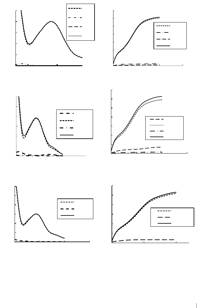
173
Acta Pharmaceutica Sciencia. Vol. 57 No. 2, 2019
Pre-processing of the UV spectral data by CAP: Signal amplification
For better assessment on signal prepossessing before PLS calibration, UV
spectra of all drugs and their mixtures (binary and ternary) were recorded at
the ratios identical to the real formulations along with CAP spectral transfor-
mation. The results are shown in Fig 3.
Pre-processing of the UV spectral data by CAP: Signal amplification
For better assessment on signal prepossessing before PLS calibration, UV spectra of all drugs and their
mixtures (binary and ternary) were recorded at the ratios identical to the real formulations along with
CAP spectral transformation. The results are shown in Fig 3.
Figure 3. UV spectra and CAP transformation of drugs and their mixtures recorded at the same ratios
as in real formulations.
In the three systems, UV spectra indicated a strong absorption signal of PAR due its higher
concentration. For PAR-PSE-CHL, PAR was added in excess than CHL and CHL by 150 and 10
0,0
0,5
1,0
1,5
2,0
2,5
200 250
Absorbance
Wavelength/nm
PAR-PSE-CHL
PAR
PSE
CHL
Mixture
0
20
40
60
80
100
120
140
200 250 300 350
CAP
Wavelength/nm
PAR-PSE-CHL
PAR
PSE
CHL
Mixture
0,0
0,5
1,0
1,5
2,0
2,5
3,0
200 250 300 350
Absorbance
Wavelength/nm
PAR-CAF-PHE
CAF
PAR
PHE
Mixture
0
20
40
60
80
100
120
140
200 250 300 350
CAP
Wavelength/nm
PAR-CAF-PHE
CAF
PAR
PHE
Mixture
0,0
0,5
1,0
1,5
2,0
2,5
3,0
200 250 300 350
Absorbance
Wavelength/nm
PAR-DPH
PAR
DPH
Mixture
0
20
40
60
80
100
120
200 250 300
CAP
Wavelength/nm
PAR-DPH
PAR
DPH
Mixture
Figure 3. UV spectra and CAP transformation of drugs and their mixtures recorded at the
same ratios as in real formulations.

174
Acta Pharmaceutica Sciencia. Vol. 57 No. 2, 2019
In the three systems, UV spectra indicated a strong absorption signal of PAR
due its higher concentration. For PAR-PSE-CHL, PAR was added in excess
than CHL and CHL by 150 and 10 times, respectively. While for PAR-CAF-
PHE, PAR was 100 and 20 times higher than PHE and CAF, respectively. For
the binary system, PAR was 20-times higher than the other drug. The high level
of PAR in the systems would explain the similarity between PAR and the spect-
rum of the mixture in all cases. The diiculty in analyzing the mixtures was
increased by the fact that the absorptivity values of CAF, CHL, PSE and DPH in
the respective formulations were much lower than the other PAR over the enti-
re spectral domain, making their accurate determination a hard-analytical task
unless the components are separated before detection. This diiculty drasti-
cally reduced the chance to apply classical spectroscopic methods which often
adopted discrete wavelengths for detection. In these cases, multivariate calib-
ration methods seem more suitable because they use simultaneously a large
number of signals per spectrum. However, considering the diiculty in solving
this type of mixtures, an appropriate pre-processing of the analytical data se-
emed necessary in order to minimize any instrumental interference and at the
same time select the most useful information to emphasize the contribution of
substances of low concentration.
As outlined earlier, CAP treatment transforms the original UV spectrum into a
new curve created by using the area underlying the spectrum. This treatment
is based on two mathematical steps as outlined earlier. Indeed, the conversion
of original UV spectra through the CAP method provided many advantages
among which the amplification of the analytical signals. Fig 3 shows the outputs
of CAP treatment of the spectral of the three systems where drugs recorded at
their actual ratios. The major dierences in comparing the original absorbance
curves with CAP plots are: a) the magnitudes on the y-axis indicated a high
amplification of the signal and this supposed to increase the sensitivity of the
quantitative measurements and help in quantifying the components even at
trace levels (like PSE, CHL, CAF and DPH in the presence of PAR), b) CAP was
able to magnify the weak signals of PSE, CHL, CAF and DPH and over the who-
le spectrum and this option is rather fundamental when applying multivariate
techniques which utilize the full spectrum to extract the maximum analytical
information, and c) Unlike UV signal, the values of CAP signal were stabilize
at constant value and this was reported for all drugs. This stable value, in fact,
is due to the additive contribution of all intensities at all wavelengths. It was
reported that CAP filtering would end up with accurate quantification of drugs
when present at trace levels
7
. The analytical matrices were built on the spectral
data. In the matrix A (dependent variables) each sample was described by n

175
Acta Pharmaceutica Sciencia. Vol. 57 No. 2, 2019
variables, corresponding to the wavelengths and to the respective absorbance
values; in the matrix C (independent variables) the samples were described
by the concentrations of each component. Two data matrices for each drug-
system were created; the first matrix contained the untreated spectral data
while the other matrix contained the spectral data upon CAP. The calibration
curves relative to the binary (PAR-DPH) and ternary systems (PAR-PSE-CHL
and PAR-CAF-PHE) along with CAP transformation are presented in Figure 4.
data. In the matrix A (dependent variables) each sample was described by n variables, corresponding
to the wavelengths and to the respective absorbance values; in the matrix C (independent variables)
the samples were described by the concentrations of each component. Two data matrices for each
drug-system were created; the first matrix contained the untreated spectral data while the other matrix
contained the spectral data upon CAP. The calibration curves relative to the binary (PAR-DPH) and
ternary systems (PAR-PSE-CHL and PAR-CAF-PHE) along with CAP transformation are presented
in Figure 4.
Figure 4. UV spectra of PAR–DPH, CAF–PAR–PHE and PAR–PSE–CHL calibration samples and
plots after CAP transformation.
0,00
0,50
1,00
1,50
2,00
2,50
200 250 300
Absorbance
Wavelength/nm
PAR-PSE-CHL
0
10
20
30
40
50
60
70
200 250 300
CAP
Wavelength/nm
PAR-PSE-CHL
0,00
0,20
0,40
0,60
0,80
1,00
200 250 300
Absorbance
Wavelength/nm
PAR-CAF-PHE
0
10
20
30
40
50
60
200 250 300
CPA
Wavelength/nm
PAR-CAF-PHE
0,00
0,50
1,00
1,50
2,00
2,50
220 240 260 280 300
Absorbance
Wavelength/nm
PAR-DPH
0
20
40
60
80
100
200 250 300
CAP
Wavelength/nm
PAR-DPH
Figure 4. UV spectra of PAR–DPH, CAF–PAR–PHE and PAR–PSE–CHL calibration samples
and plots after CAP transformation.

176
Acta Pharmaceutica Sciencia. Vol. 57 No. 2, 2019
Determination of pharmaceuticals in binary and ternary
drugs systems by PLS
PLS and CAP-PLS can be created and applied to predict drugs without previ-
ous knowledge on calibrated solutes. However, intense overlapping between
drugs and strong inuence of excipients would aect the prediction power of
PLS
15,16
. The performance of before and after signal filtering drugs prediction
in validation set is provided in Table 3.
Table 3. External prediction of drugs in different systems.
Drug
PLS
a
CAP-PLS
b
PRESS REP% Rec% PRESS REP% Rec%
PAR-PSE-CHL
PAR 0.32 3.9 96.3 (4) 0.06 1.7 98.0 (3)
PSE 2.95 12.6 95.2 (5) 0.13 3.1 97.7 (4)
CHL 2.32 11.2 94.0 (5) 0.15 2.5 97.7 (4)
PAR-CAF-PHE
PAR 3.41 14.0 106.8 (5) 0.82 5.8 99.2 (3)
CAF 2.93 10.0 97.4 (4) 0.45 4.3 103.6 (3)
PHE 1.54 8.0 95.8 (4) 0.18 2.1 101.4 (3)
PAR-DPH
PAR 0.45 3.4 96.9 (3) 0.14 2.1 98.5 (2)
DPH 0.64 4.9 95.7 (5) 0.19 3.0 97.0 (4)
a
Spectral range 200-300 nm (101 point/spectrum). Calibration and prediction steps were
carried out using Eqs1-3. Calibration and validation sets are provided in Table 1. PLS variables
were provided between brackets for each system.
b
Prior to PLS calibration, spectral data were ltered using cumulative area pre-processing
method (Eqs 11&12) and PLS variables were provided between brackets for each system.
As shown in Table 3, PLS was workable to predict PAR-PSE-CHL and the best
prediction observed for PAR with a REP value 3.9%. In general, PLS was not
so-eective for predicting PSE in the mixtures with a high REP 12.6% which
may not acceptable for pharmaceutical analysis. The modest prediction of PSE
was mainly attributed to the intense spectral-overlapping with other drugs
(Table 2). Accordingly, application of PLS for PSE and CHL may not be wor-
kable taking into account the negative inuence of excipients in real cases. As
indicated in Table 3, the performance of CAP-PLS was very promising with

177
Acta Pharmaceutica Sciencia. Vol. 57 No. 2, 2019
REP values of 1.7%, 2.5%, and 3.1% for PAR, CHL, and PSE, respectively, and
this reected the high closeness between nominal and predicted values. The
earlier results showed that CAP-PLS outperformed PLS for predicting PSE and
CHL in ternary systems. In general, CAP-PLS was excellent for predicting all
drugs with PRESS values less than 1.0 in all cases. The same results were noted
for PAR-CAF-PHE where CAP-PLS outperformed PLS for drugs quantificati-
on with final REP 2.1-5.8%. The best prediction was reported for PHE down
to 1.0 mg/L in mixtures rich with PAR and CAF. Due to the lesser spectral
overlapping, both PLA and CAP-PLS were both of comparable performance
for PAR-DPH quantification, REP was all lower than 5% for both methods.
The superiority of CAP-PLS was mainly attribute to the eective signal filtering
prior to PLS calibration which improved final prediction
7
.
Quantification of drugs in real formulations
The real testing of the proposed CAP-PLS method will be assessed by analyzing
binary and ternary systems in the commercial formulations. Direct analysis of
drugs in commercial tablets is not an easy task due to the presence of drugs
in unequal quantities and also the presence of excipients that did not involve
while constructing calibration mixtures. In addition to the above, the drugs
are often placed at concentrations far higher than the calibration solutions.
Therefore, all the ingredients should be properly diluted to be measured with
acceptable sensitivity. In this case, signal filtering/enhancing will be essential
before running classical PLS calibration. It is rather essential to mention that
matrix-cleaning methods may be adopted to eliminate/reduce the inuence
of interferences before running analysis
22-24
. It was necessary to evaluate the
eect of dilutions on drugs quantification. The spectral shapes of the systems
and at variable dilutions are provided in Figure 5. Drugs quantification in real
formulations is provided in Table 4 and the PLS outputs are provided for com-
parison purposes.

178
Acta Pharmaceutica Sciencia. Vol. 57 No. 2, 2019
Figure 5. UV spectra of marketed formulations after extraction and dilution with distilled water and
CAP plots.
0,00
0,50
1,00
1,50
2,00
2,50
3,00
200 250 300
Absorbance
Wavelength/nm
PAR-PSE-CHL
DF 1:30
DF 1:25
DF 1:15
DF 1:10
0
20
40
60
80
100
120
200 220 240 260 280 300
CAP
Wavelength/nm
PAR-PSE-CHL
DF 1:30
DF 1:25
DF 1:15
DF 1:10
0,00
0,50
1,00
1,50
2,00
2,50
3,00
3,50
200 220 240 260 280 300
Absorbance
Wavelength/nm
PAR-CAF-PHE
DF 10
DF 15
DF 20
DF 40
0
50
100
150
200
250
200 220 240 260 280 300
CAP
Wavelength/nm
PAR-CAF-PHE
DF 10
DF 15
DF 20
DF 40
0,00
0,20
0,40
0,60
0,80
1,00
1,20
1,40
220 240 260 280 300
Absorbance
Wavelength/nm
PAR-DPH
DF 10
DF 15
DF 25
DF 50
0
10
20
30
40
50
60
70
80
200 220 240 260 280 300
CAP
Wavelength/nm
PAR-DPH
DF 10
DF 15
DF 25
DF 50
Figure 5. UV spectra of marketed formulations after extraction and dilution with distilled
water and CAP plots.

179
Acta Pharmaceutica Sciencia. Vol. 57 No. 2, 2019
Table 4. Implementation of PLS and CAP-PLS for drugs quantication on different systems.
System
Claimed
(mg/tablet)
PLS CAP-PLS
Predicted
(mg/tablet)
a
Mean
Recovery
% (n=3)
Precision
(n=3, RSD%)
Predicted
(mg/tablet)
Mean
Recovery
% (n=3)
Precision
(n=3, RSD%)
PAR-CAF-PHE
PAR 500 503.0 100.6 8.4 501.0 102.6 2.1
CAF 25 26.0 104.0 6.5 24.7 103.0 3.1
PHE 5 4.5 90.0 7.3 5.1 94.3 4.2
PAR-PSE-CHL
PAR 300 508.0 99.0 5.0 504.0 98.9 3.1
PSE 30 28.7 102.0 7.0 25.2 104.6 2.7
CHL 2 1.8 99.0 4.6 2.1 99.0 1.5
PAR-DPH
PAR 500 503 101.2 2.1 502 104.2 0.8
DPH 25 28 105.3 3.1 24 106.3 1.0
As indicated in Fig 5, identical UV spectra and CAP signals were observed for
the systems. Higher intensities (for CAP signals) were observed at lower dilu-
tions (DF 10 and 15). Initial analysis indicated that running PLS or CAP-PLS
using signals recorded at DF 10 or 15 ended up with high prediction errors. In
fact, at lower DF the content of drugs is much higher than those used in ca-
libration mixtures making external prediction is not valid. As already known,
the external prediction of solutes is often dependent on the ranges selected
in the calibration mixtures. In the current systems, the best prediction in real
formulations was achieved at DF 15, 20, and 25 for PAR-DPH, PAR-CAF-PHE,
and PAR-PSE-CHL, respectively. Both models were used to assay drugs in real
formulations which based the same composition of the calibration mixtures.
The results, provided in Table 4, indicated the high performance of CAP-PLS
for accurate quantification of drugs in binary and ternary systems. For ter-
nary mixtures, CAP-PLS manifested better accuracy than PLS with recoveries
94.3%-102.6% and precisions (RSD) 2.1-3.1% for PAR-CAF-PHE and 98.9%-
104.6% and 1.5-3.1 for PAR-PSE-CHL. It seems that signal filtering by CAP has
passively reected on the final PLS performance. For the other binary system,
a good agreement was observed between experimental and nominal levels (ob-
tained by both models) of the commercial formulation. Again, PLS calibration
created using CAP-filtered-data generated the best results and may properly
handed the negative inuence of excipients present in the dosage forms.

180
Acta Pharmaceutica Sciencia. Vol. 57 No. 2, 2019
UV-signal filtering by CAP prior to PLS was found more eective than PLS
when handling ternary drug mixtures. The proposed CAP-PLS can handle
systems in which one component present at lower levels in the formulation.
CAP simply estimated the cumulative sum of the areas under curve between
two consecutive wavelengths which improved the analytical signal. The pro-
posed CAP-PLS was eective to predict two ternary systems (PAR-CAF-PHE
and PAR-PSE-CHL) of intense overlapping and containing one ingredient ava-
ilable at lower concentration. For binary system, application of CAP-PLS was
as good as classical PLS along with raw UV signals. CAP, as an appropriate
pretreatment method, have been succeeded for accurate and simultaneous
quantification of commercial pharmaceutical formulations in which the ratio
between the components is highly variable.
ACKNOWLEDGEMENTS
The authors thank Deanship of Graduate Studies (The Hashemite University,
Jordan) and for the financial support to this research. The researchers also
thank all the technical stas at chemistry department for their continued help.
List of abbreviations
CAF Caeine
CHL Chlorpheniramine Maleate
CAP Cumulative Area Pre-Processing
DPH Diphenhydramine Hydrochloride
LOD Limit of Detection
LOQ Limit of Quantification
NAS Net Analyte Signal
PLS Partial Least Squares
PRESS Prediction Error Sum of Square
PSE Pseudoephedrine Hydrochloride
REP% Relative Error of Prediction
SEL Selectivity
SEN Sensitivity

181
Acta Pharmaceutica Sciencia. Vol. 57 No. 2, 2019
REFERENCES
1. Shaikh, K.; Devkhile, A. Simultaneous determination of aceclofenac, paracetamol, and
chlorzoxazone by RP-HPLC in pharmaceutical dosage form. J. Chroma. Sci. 2008, 46, 649-
652.
2. Ali, N.; Zaazaa, H.; Abedelkawy, M.; Magdy, M. Simultaneous determination of paraceta-
mol and diphenhydramine hydrochloride in presence of paracetamol degradation product. J.
Pharm. Anal. Acta. 2011, 2, 9.
3. Dou, Y.; Sun, Y.; Ren, Y.; Ren, Y. Artificial neural network for simultaneous determination
of two components of compound paracetamol and diphenhydramine hydrochloride powder
on NIR spectroscopy. Anal. Chim. Acta. 2005, 528, 55-61.
4. Goicoechea, H.; Olivieri, A. Simultaneous multivariate spectrophotometric analysis of pa-
racetamol and minor components (diphenhydramine phenylpropanolamine) in tablet prepa-
rations. J. Pharm. Biomed. Anal. 1999, 20. 255- 261.
5. Hegazy, M.; Abbas, S.; Zaazaa, H. Resolution of overlapped quaternary spectral bands by
net analyte signal based methods; an application to dierent combinations in tablets and
capsules. J. Anal. Chem. 2015, 70, 450–458.
6. De Luca, M.; Ioele, G.; Spatari, C.; Ragno, G. Optimization of wavelength range and data
interval in chemometric analysis of complex pharmaceutical mixtures. J. Pharm. Anal. 2016,
6, 64-69.
7. De Luca, M.; Ioele, G.; Ragno, G. Cumulative area pre-processing (CAP): A new treatment
of UV data for the analysis of complex pharmaceutical mixtures. J. Pharm. Biomed. Anal.
2014, 90, 45– 51.
8. Singh, V.; Daharwal, S.; Suresh, P. A review of instrumental analytical methods to assay
active ingredients in multicomponent pharmaceutical formulations. Columb. J. Pharm. Sci.
2014, 1, 27-39.
9. Qi, M.; Wang, P.; Zhou, L.; Sun, Y. Simultaneous determination of four active components
in a compound formulation by liquid chromatography. Chromatographia. 2003, 58, 183-
186.
10. Belal, T.; Abdel-Hay, K.; Clark, R. Selective determination of dimenhydrinate in presence
of six of its related substances and potential impurities using a direct GC/MS method. J. Adv.
Res. 2016, 7, 53-58.
11. Gergov, G.; Alin, A.; Doychinova, M.; De Luca, M.; Simeonov, V.; Al-Degs, Y. Assessment
of dierent PLS algorithms for quantification of three spectrally overlapping drugs. Bull.
Chem. Comm. 2017, 49, 410–421.
12. Khajehsharifi, H.; Sadeghi, M.; Pourbasheer, E. Spectrophotometric simultaneous deter-
mination of ceratine, creatinine, and uric acid in real samples by orthogonal signal correcti-
on–partial least squares regression. Chemical Monthly. 2009, 140, 685–691.
13. Gabrielsson, J.; Jonsson, H.; Airiau, C.; Schmidt, B.; Escott, R.; Trygg, J. OPLS method-
ology for analysis of pre-processing eects on spectroscopic data. Chemom. Intell. Lab. Syst.
2006, 84, 153–158.
14. Svensson, O.; Kourti, T.; MacGregor, J. An investigation of orthogonal signal correction
algorithms and their characteristics. J. Chemomet. 2002, 16, 176–188

182
Acta Pharmaceutica Sciencia. Vol. 57 No. 2, 2019
15. Al-Degs, Y.; El-Sheik, A.; Issa, A.; Al-Ghouti, M.; Sunjuk, M. Asimple and accurate analy-
tical method for determination of three commercial dyes in dierent water systems using
partial least squares regression. Wat. Sci. Technol. 2012, 66, 1647-1655.
16. Goicoechea, H.; Olivieri, A. Simultaneous determination of phenobarbital and phenyto-
in in tablet preparations by multivariate spectrophotometric calibration. Talanta. 1998, 47,
103-108
17. Haaland, D.; Thomas, E. Partial least-squares methods for spectral analyses.1. Relation to
other quantitative calibration methods and the extraction of qualitative information. Anal.
Chem. 1988, 60, 1193-1202.
18. Andersson, M. A comparison of nine PLS1 algorithms. J. Chemomet. 2009, 23, 518-529
19. Noor, P.; Khanmohammadi, M.; Roozbehani, B.; Garmarudi, A. Evaluation of ATR-FTIR
spectrometry in the fingerprint region combined with chemometrics for simultaneous deter-
mination of benzene, toluene, and xylenes in complex hydrocarbon mixtures. Chem. Month.
2018, 149, 1341-1347.
20. Lorber, A.; Faber, K.; Kowalski, B. Net analyte signal calculation in multivariate calibrati-
on. Anal. Chem. 1997, 69, 1620-1626.
21. Yousefinejad, S.; Hemmateenejad, B. Simultaneous spectrophotometric determination of
paracetamol and para-aminophenol in pharmaceutical dosage forms using two novel multi-
variate standard addition methods based on net analyte signal and rank annihilation factor
analysis. Drug test. Anal. 2012, 4, 507-514.
22. El-Sheikh, A; Al-Jafari, M; Sweileh, J. Solid phase extraction and uptake properties of
multi-walled carbon nanotubes of dierent dimensions towards some nitro-phenols and
chloro-phenols from water, Int. J. Env. Anal. Chem. 2012, 92, 190-209.
23. El-Sheikh, A; Sweileh, J; Saleh, M. Partially-pyrolyzed olive pomace sorbent of high per-
meability for preconcentration of metals from environmental waters, J. Hazard. Mat. 2009,
169, 58-64.
24. Al-Hashimi, N; Aleih, H; Fasfous, I; AlKhatib, H. Multi-Walled Carbon Nanotubes as Ef-
ficient Sorbent for the Solid Bar Microextraction of non-Steroidal Anti-Inammatory Drugs
from Human Urine Samples. Curr. Pharm. Anal. 2018, 14, 239-246.

183
Acta Pharmaceutica Sciencia. Vol. 57 No. 2, 2019
Acta Pharm. Sci. Vol 57 No: 2. 2019
DOI: 10.23893/1307-2080.APS.05711
Chemical Composition and Comparative
Antibacterial Properties of Basil Essential
Oil against Clinical and Standard Strains of
Campylobacter spp.
Aysegul Mutlu-Ingok
1,2*
, Burcu Firtin
1
, Funda Karbancioglu-Guler
1
1 Istanbul Technical University, Faculty of Chemical and Metallurgical Engineering, Department of Food Engineering,
Istanbul, Turkey.
2 Duzce University, Akcakoca Vocational School, Food Technology Department, Duzce, Turkey.
INTRODUCTION
Campylobacter spp. is considered to be the most common bacterial cause of
human gastroenteritis in the world
1
. Food-borne Campylobacter infections
are considered to be caused by animal origin foods, mainly poultry and po-
ultry products. Besides poultry, raw milk, pork, beef, lamb and seafood are
responsible of Campylobacter infections
2
. The antimicrobial resistance of
ABSTRACT
This study has aimed to evaluate comparative antibacterial activity of basil essenti-
al oil against clinical and standard isolates of Campylobacter spp. by dierent met-
hods as agar well diusion, agar and broth dilution methods. Gas Chromatography
(GC) and Gas Chromatography/Mass Spectrometry (GC/MS) analysis were also
examined to determine the chemical composition of the tested essential oil. GC/
MS analysis showed that, basil essential oil was predominated by methyl chavicol
(86.6%) followed by 1,8-cineole (2.8%) and α-bergamotene (2.4%). Although, in-
hibition zone diameters were in the range of 10.9±0.8 to 21.8±1.4 mm, higher MIC
values were obtained against clinical strains compared with standard ones. Due to
the dierences in antimicrobial resistance of the clinical and standard strains, an-
timicrobial activity tests should be carried out with isolates from dierent sources.
Keywords: Basil essential oil, Chemical composition, Agar well diusion, Broth
microdilution, Agar dilution.
*Corresponding Author: Aysegul Mutlu-Ingok, e-mail: aysegulmutlu@duzce.edu.tr
Aysegul Mutlu-Ingok ORCID Number: 0000-0001-9571-0053
Burcu Firtin ORCID Number: 0000-0002-4633-9582
Funda Karbancioglu-Guler ORCID Number: 0000-0001-6576-0084
(Received 15 January 2019, accepted 23 February 2019)

184
Acta Pharmaceutica Sciencia. Vol. 57 No. 2, 2019
thermophilic campylobacters, including Campylobacter jejuni and C. coli has
been identified especially to tetracyclines and uoroquinolones at important
levels in many dierent parts of the world
1,3
. Using chemical compounds have
limits because of their carcinogenic eects, acute toxicity, and environmental
hazard potential
4
. Increasing resistance to currently used antimicrobials and
consumer concerns about using chemical preservatives lead to investigation
of alternative strategies to prevent and control these microorganisms. Despite
the high number of studies on the antimicrobial eects of essential oils (EOs),
most studies have focused on pathogenic bacteria like Staphylococcus aureus,
Escherichia coli, and Bacillus cereus
5
.
Essential oils which were synthesized naturally in dierent plant parts are
complex volatile compounds. They can be extracted from medicinal aromatic
plants and have strong antimicrobial activity against various bacterial, fungal,
and viral pathogens. In addition to their antibacterial properties, they have
antiviral, antimycotic, antitoxigenic, antiparasitic, insecticidal, antimutageni-
city, cytoprotective, moderation of insulin secretion analgesic, neuroprotecti-
ve, antioxidant, antiproliferative proapoptotic anxiolytic-like activities
6
. Their
wide range of antimicrobial activity was a result of dierent types of aldehydes,
phenolics, terpenes, and other antimicrobial compounds
4
. Mechanism of an-
timicrobial action is still lacking although a few studies have been elucidated
7
.
Basil is the common name for the culinary herb Ocimum basilicum of the fa-
mily Lamiaceae (Labiatae). Although the basil essential oil’s antibacterial acti-
vity is associated with its high content in linalool and estragole, antimicrobial
spectrum is restricted to specific bacteria other than Campylobacter spp.
8
Alt-
hough in few studies antimicrobial activity of basil essential oil against Camp-
ylobacter spp. has been mentioned
9,10
, our literature review revealed that the
dierences of antimicrobial eects against clinical and standard Campylobac-
ter isolates were not discussed.
In this study, it was aimed to evaluate comparative antibacterial activity of ba-
sil essential oil against clinical and standard isolates of Campylobacter jejuni
and Campylobacter coli by dierent methods as agar well diusion, agar and
broth dilution methods. Gas Chromatography (GC) and Gas Chromatography/
Mass Spectrometry (GC/MS) analyses also examined the chemical compositi-
on of the tested EO.
METHODOLOGY
Bacterial culture and essential oils
The antimicrobial activity of the cold pressed basil essential oil was tested

185
Acta Pharmaceutica Sciencia. Vol. 57 No. 2, 2019
against clinical Campylobacter jejuni, Campylobacter coli identified by Mat-
rix- Assisted Laser Desorption/Ionization time- of- ight Mass Spectrometry
(MALDI TOF MS)
11
and standard Campylobacter jejuni (ATCC 33660), Camp-
ylobacter coli (NCTC 12525). Basil essential oil was obtained in food grade
form from “International Flavors & Fragrances (IFF)”, Gebze, Kocaeli (Tur-
key). Dilutions were made in 10% dimethyl sulfoxide (DMSO, Merck). Before
analysis, basil essential oil was sterilized by filtration through 0.22 μm filters
(Minisart
®
Syringe Filter, Sartorius Stedim Biotech GmbH, Germany) and sto-
red in dark at 4 °C.
Gas Chromatography (GC)
Essential oils were analyzed by GC-FID using an Agilent 7890B GC (Agilent,
Palo Alto, CA) with a ame ionization detector (FID). The chromatographic
separation was accomplished using an Agilent HP- Innowax column (60 m x
0.25 mm Ø, with 0.25 μm film thickness) with a helium as a carrier gas (0.7
mL/ minute). GC oven temperature was kept at 60 °C for 10 min and program-
med to 220 °C at a rate of 4 °C/ minute and then kept constant at 220 °C for
10 min and programmed to 240 °C at a rate of 1 °C/ minute. The injector and
ame ionization detector temperatures were adjusted to 250 °C. The relative
percentage amounts of the separated compounds were calculated from FID
chromatograms.
Gas Chromatography-Mass Spectrometry (GC/MS)
The essential oils were analyzed by GC/MS using an Agilent 7890B GC coupled
with a 5977B MSD (Agilent, Palo Alto, CA). The same column and analytical
conditions were used for both GC/MS and GC/FID. The mass range was recor-
ded from m/z 35 to 425. The injector temperature was adjusted to 250 °C. MS
were recorded at 70 eV. Alkanes were used as reference points in the calculati-
on of relative retention indices (RRI). The components of EOs were identified
by using Wiley 9- Nist 11 Mass Spectral Database and standard Alkan series
(C7-C40).
Agar-well diusion assay
Inhibition zone diameters were determined using previously described method
with slight modifications
12
. Bacterial inoculum was prepared in Mueller-Hin-
ton Broth (MHB, Merck, Darmsdat, Germany) for standard isolate and MHB
with 5% horse blood for clinical isolate and incubated at 42 °C for 48 h un-
der microaerophilic conditions created by Anaerocult® C (Merck, Darmsdat,
Germany). Concentrations of bacterial suspensions were adjusted to approxi-
mately 10
8
cfu/mL and 100 μL of culture suspension was spreaded on Camp-

186
Acta Pharmaceutica Sciencia. Vol. 57 No. 2, 2019
ylobacter Blood-Free Selective Agar Base medium (modified CCDA, Merck,
Darmsdat, Germany) for standard isolate, Mueller-Hinton Agar (MHA, Merck,
Darmsdat, Germany) medium with 5% horse blood for clinical isolate. Three
wells were cut out of agar and filled with 5 μL, 10 μL and 20 μL of basil EO.
The inoculated plates were incubated at 42 °C for 48 h under microaerophi-
lic conditions. After incubation, inhibition zone diameters were measured. All
experiments were performed in triplicate. Zones of inhibition (including the
6 mm of the well) were expressed as mean values with ± standard deviation.
Broth microdilution assay
Broth microdilution method was used to determine the minimum inhibition
concentrations (MICs), which was described previously by Wiegand et al.
13
.
Stock solution was prepared in 10% DMSO and two-fold serial dilutions of EO
were prepared. After sub-culturing in MHB, bacterial concentration was adjus-
ted to approximately 10
8
cfu/mL. The 96-well plates were prepared by dispen-
sing, into each well, 95 μL of MHB, 100 μL of EO and 5 μL of the inoculants.
The final volume in each well was 200 μL. The microplates were incubated at
42 °C for 24 h under microaerophilic conditions. MIC values were determined
spectrophotometrically by measuring the optical density at an absorbance of
600 nm (Synergy HT, BioTek Instruments Inc., Winooski, VT, USA). Negative
controls (involving 195 μL of MHB and 5 μL of inoculum but no EO) for each
microorganism and sterility controls (involving 100 μL MHB and 100 μL EO
but no inoculum) for each EO concentrations were prepared.
Agar dilution method
For clinical strains, to determine minimum inhibitory concentrations (MICs),
agar dilution method according to Stepanović et al.
14
was used with slight mo-
difications. This method based on preparation of MHA with 5% horse blood
with the additions of 1% Tween 20 and dierent concentration of essential oils
after sterilization of agar. Test plates were prepared with 19 mL of MHA, and 1
mL of two-fold dilutions of essential oils. After adjusting bacterial concentrati-
on approximately to 10
8
cfu/mL, 10 μL of culture suspension was inoculated to
agar plates. Plates were incubated for 48 h at 42 °C in microaerophilic condi-
tions. The MICs were defined as the lowest concentration of essential oils that
inhibited visible growth of microorganisms.
14
RESULTS AND DISCUSSION
The chemical composition of the basil EO determined with GC/MS is given in
Table 1. The main compound identified in the basil essential oil was methyl
chavicol (86.6%). These results are consistent with those reported in the lite-

187
Acta Pharmaceutica Sciencia. Vol. 57 No. 2, 2019
rature. Dierences of constituents and their amounts may be related with the
geographical origin of the plant, dierent parts of plants, extraction method
and season of harvest
15
.
Table 1. Chemical compositions of basil essential oil.
No Compound RI
a
Peak area (%)
b
1 1,8-Cineole 1220 2.8
2
α-Bergamotene
1605 2.4
3 Methyl chavicol 1701 86.6
Total 91.8
a
: Retention index was calculated for all volatile constituents using a homologous series of
n-alkanes C7- C40, b: Peak area obtained by GC-FID.
Although dierent chemical profiles of basil essential oil were reported in lite-
rature, methyl chavicol with high citral contents (methyl chavicol/citral) was
previously detected as a ‘‘new chemo type’’ in the Turkish basils
16
. In additi-
on to geological origin, chemical constituents varied with dierent seasons
17
.
Generally, the chemical composition profile of basil essential oil confirms
previous studies. Methyl chavicol was reported as major constituent in India
(78.3%)
18
. In another study, three chemotypes of Ocimum basilicum (O. basi-
licum) were identified as a major methyl chavicol-rich type (>65%), a methyl
chavicol (55%)-linalool (20–30%) type, a linalool (42–45%) and eugenol (15%)
type
19
. For this respect, O. basilicum used in this study was in methyl chavicol-
rich type with 86.6% methyl chavicol. High content of methyl chavicol was also
confirmed by Vieira and Simon
20
with 47% methyl chavicol content.
The inhibition zone diameters measured ranged from 12.3±1.6 to 21.8±1.4 mm
and 10.9±0.8 to 20.4±2.4 mm for clinical and standard Campylobacter iso-
lates, respectively (Table 2). Considering the all results, mean inhibiton zone
diameter was 15.94±1.55 mm. Similar to current study, mean zone diameters
were reported as 12.48 mm and 13.2 mm against gram positive and gram ne-
gative bacteria, respectively
21
. Smaller inhibition zones were also reported by
Predoi et al.
22
as 7-10 mm against Escherichia coli, Staphylococcus aureus
and methicillin-resistant Staphylococcus aureus. In literature, inhibition zone
diameters were varied depending on dierent extracts. It was reported that
although methanol extracts showed inhibition zones against Pseudomonas ae-
ruginosa, Shigella sp., Listeria monocytogenes, Staphylococcus aureus and
two dierent strains of Escherichia coli, chloroform and acetone extracts of O.
basilucum had no eect
23
.

188
Acta Pharmaceutica Sciencia. Vol. 57 No. 2, 2019
Poor solubility and high volatility of essential oils limit the usage of diusion
tests. It is suggested to use agar or broth dilution methods for true antimicro-
bial activity evaluation
24
. With this respect, in this study, in vitro antimicrobial
activity of basil essential oil was not tested only by agar diusion method but
also dilution methods against clinical and standard isolates of Campylobac-
ter spp. (Table 2). It was reported that both agar dilution and broth microdi-
lution methods were equally suitable against Campylobacter spp. and highly
correlated
25
. In this study, since broth microdilution method did not give any
results against clinical strains, agar dilution method was used by taking this
perspective into consideration. Tested essential oil displayed varying degree
of antibacterial activity with MIC values ranging from 105.16 to 1787.7 μg/mL.
Interestingly, MIC values against clinical ones were higher than standard iso-
lates. Higher MIC values indicate that clinical strains are more resistant than
standard strains against basil essential oil.
Table 2. Antimicrobial activity of basil essential oil against Campylobacter spp.
Isolate
Amount (µL) MIC (µg/mL)
5 µL 10 µL 20 µL
Inhibition zone diameter (mm)
C. jejuni (Clinical)
Streptomycin
b
NA
a
38.0±1.8
13.0±2.2
40.0±2.4
20.1±1.6
45.0±1.4
1787.7
NT
c
C.coli (Clinical)
Streptomycin
NA
32.0±1.4
12.3±1.6
38.0±1.4
21.8±1.4
40.0±1.3
889.08
NT
C. jejuni (ATCC 33560)
Streptomycin
NA
21.3±2.1
10.9±0.8
24.7±1.5
20.4±2.4
30.0±1.3
105.16
NT
C. coli (NCTC 12525)
Streptomycin
NA
21.7±1.6
11.8±1.1
24.8±0.7
17.2±1.3
28.8±1.3
219.88
NT
a
NA: No activity,
b
: Standard antibiotic,
c
: Not tested
Although the same MIC values were reported for essential oils against dierent
strains in literatue, in this study dierences in MIC values were found against
the clinical and standard strains of Campylobacter. In literature, dierent MIC
values were reported. Antibacterial and antifungal activities of essential oils of
twelve Ocimum basilicum L. cultivars which were grown in Serbia were inves-
tigated by Beatovic et al.
26
. However, lower MIC values than current study were
reported, they were ranging from 0.009-11.74 μg/mL. Silveira et al.
27
, reported
MIC values from 0.075 to 2.5 μg/mL against S. aureus, L. monocytogenes, B.
cereus, Yersinia enterocolitica, E. coli and S. typhimurium. In another study,
mean MIC values were detected as 0.75 and 0.73 μg/mL against 6 gram positive
and 12 gram negative bacteria respectively
21
. Higher MIC values were also repor-

189
Acta Pharmaceutica Sciencia. Vol. 57 No. 2, 2019
ted for gram positive bacteria as 18-36 μg/mL, and for gram-negative bacteria
as 9-18 μg/mL
28
. By the existence of dierent EO components with respect to
harvesting season dierences as well as extraction method, dierent antimicro-
bial activity levels can be obtained. These dierences may be due to this fact
15
.
Antimicrobial spectrum of basil essential oil was reported as restricted to spe-
cific bacteria as Staphylococcus spp., Enterococcus spp., E. coli, P. aerugino-
sa, Acinetobacter baumannii, Aeromonas hydrophila, B. cereus, Bacillus sub-
tilis, Enterobacter spp., Listeria spp., Proteus spp., Salmonella spp., Serratia
marcescens, and Y. enterocolitica and fungi as Candida spp., Rhodotorula
spp., and Saccharomyces cerevisae
8
. Although basil essential oil has restricted
antimicrobial activity, in current study it has also been proven that it has anti-
microbial activity against Campylobacter spp.
This study described antibacterial eiciency dierences of basil essential oil
against clinical and standard isolates of Campylobacter spp., as well as the
chemical composition of corresponding essential oil. The results indicated that
tested EO has varying degree of antibacterial eiciency against both C. jejuni
and C. coli isolates. However, with in vitro experiments, in vivo studies are
also required because antimicrobial eect showed dierences even between
clinical and standard strains. In addition, optimum essential oil concentration
should be determined to ensure antimicrobial activity and acceptable sensorial
properties.
ACKNOWLEDGEMENTS
The authors wish to thank Anadolu University, Medicinal Plants, Drugs and
Scientific Research Center for GC and GC-MS analyses of basil essential oil.
This research was supported by Istanbul Technical University, Scientific Rese-
arch Projects (Project no, 38819).
REFERENCES
1. WHO. The Global View of Campylobacteriosis, Report of an expert Consultation. Utrecht,
Netherlands, 2002.
2. Nachamkin, I.; Szymanski, C. M.; Blaser M. J. Campylobacter (No. Ed. 3). ASM Press.
ProQuest Ebook Central, 2008, 54.
3. Moore J. E; Barton, M. D., Blair, I. S., Corcoran, D., Dooley, J. S., Fanning S., Kempf, I.;
Lastovica, A. J.; Lowery, C. J.; Matsuda, M.; McDowell, D.A.; McMahon, A.; Millar, B. C.;
Rao, J. R.; Rooney, P. J.; Seal, B. S.; Snelling, W. J.; Tolba, O. The epidemiology of antibiotic
resistance in Campylobacter. Microbes Infect, 2006, 8, 1955-1966.
4. Swamy, M. K.; Akhtar, M. S.; Sinniah, U. R. Antimicrobial properties of plant essential
oils against human pathogens and their mode of action: an updated review. Evid Based
Complement Alternat Med, 2016, 3012462.

190
Acta Pharmaceutica Sciencia. Vol. 57 No. 2, 2019
5. Mutlu-Ingok, A.; Karbancioglu-Guler, F. Cardamom, Cumin, and Dill Weed Essential
Oils: Chemical Compositions, Antimicrobial Activities, and Mechanisms of Action against
Campylobacter spp. Molecules, 2017, 22, 1191.
6. Joshi, R. K. Role of Natural Products against Microorganisms. Am J Clin Microbiol
Antimicrob., 2018, 1, 1005.
7. Chouhan, S.; Sharma, K.; Guleria, S. Antimicrobial Activity of Some Essential Oils—
Present Status and Future Perspectives. Medicines, 2017, 4, 58.
8. Sakkas, H.; Papadopoulou, C. Antimicrobial Activity of Basil, Oregano, and Thyme
Essential Oils. J Microbiol Biotechnol, 2017, 28, 429-438.
9. Smith-Palmer, A.; Stewart, J.; Fyfe, L. Antimicrobial properties of plant essential oils
and essences against five important food-borne pathogens. Lett Appl Microbiol, 1998, 26,
118–122.
10. Friedman, M.; Henika, P. R.; Mandrell, R. E. Bactericidal activities of plant essential
oils and some of their isolated constituents against Campylobacter jejuni, Escherichia coli,
Listeria monocytogenes, and Salmonella enterica. J Food Protect, 2002, 65, 1545–1560.
11. Şamlı, A.; Ayaş, R.; Ülger, N. Akut bakteriyel gastroenterit etkenleri arasında
Campylobacter turlerinin yeri ve antibiyotiklere duyarlılıkları. ‘Ulusal Klinik Mkrobiyoloji
Kongresi, 2013, 408.
12. Deans, S.; Ritchie, G. Antimicrobial properties of plant essential oils. Int J Food Microbiol,
1987, 5, 165-180.
13. Wiegand, I.; Hilpert, K.; Hancock, R. E. W. Agar and broth dilution methods to determine
the minimal inhibitory concentration (MIC) of antimicrobial substances. Nat. Protoc. 2008,
3, 163–175.
14. Stepanović, S.; Antić, N.; Dakić, I.; Švabić-Vlahović, M. In vitro antimicrobial activity of
propolis and synergism between propolis and antimicrobial drugs. Microbiol Res, 2003, 158,
353-357.
15. Burt, S. Essential oils: Their antibacterial properties and potential applications in
foods—a review. Int J Food Microbiol, 2004, 94, 223–253.
16. Telci, I.; Bayram, E.; Yılmaz, G.; Avcı, B. Variability in essential oil composition of Turkish
basils (Ocimum basilicum L.). Biochem. Syst. Ecol., 2006, 34, 489-497.
17. Hussain, A. I.; Anwar, F.; Sherazi, S. T. H.; Przybylski, R. Chemical composition,
antioxidant and antimicrobial activities of basil (Ocimum basilicum) essential oils depends
on seasonal variations. Food Chem., 2008, 108, 986-995.
18. Chowdhury, J. U.; Bhuiyan, M. N. H.; Saha, G. C.; Nada, K.; Rahim, M. Compositions of
the essential oils from two types of ocimum basilicum introduced in Bangladesh. Bangladesh
J Sci Ind Res, 2013, 48, 217-220.
19. Yayi, E.; Moudachirou, M.; Chalchat, J. C. Chemotyping of three Ocimum species from
Benin: O. basilicum, O. canum and O. gratissimum. J Essent Oil Res., 2001, 13, 13-17.
20. Vieira, R. F.; Simon, J. E. Chemical characterization of basil (Ocimum spp.) found in the
markets and used in traditional medicine in Brazil. Econ Bot., 2000, 54, 207-216.
21. Gaio, I.; Saggiorato, A. G.; Treichel, H.; Cichoski, A. J.; Astolfi, V.; Cardoso, R. I.; ... &

191
Acta Pharmaceutica Sciencia. Vol. 57 No. 2, 2019
Cansian, R. L. Antibacterial activity of basil essential oil (Ocimum basilicum L.) in Italian-
type sausage. J Verbrauch Lebensm, 2015, 10, 323-329.
22. Predoi, D.; Iconaru, S. L.; Buton, N.; Badea, M. L.; Marutescu, L. Antimicrobial
Activity of New Materials Based on Lavender and Basil Essential Oils and Hydroxyapatite.
Nanomaterials, 2018, 8, 291.
23. Kaya, I.; Yigit, N.; Benli, M. Antimicrobial activity of various extracts of Ocimum
basilicum L. and observation of the inhibition eect on bacterial cells by use of scanning
electron microscopy. Afr J Tradit Complement Altern Med., 2008, 5, 363-369.
24. Suppakul, P.; Miltz, J.; Sonneveld, K.; Bigger, S. W. Antimicrobial properties of basil and
its possible application in food packaging. J. Agric. Food Chem., 2003, 51, 3197-3207.
25. Luber, P.; Bartelt, E.; Genschow, E.; Wagner, J.; Hahn, H. Comparison of broth
microdilution, E Test, and agar dilution methods for antibiotic susceptibility testing of
Campylobacter jejuni and Campylobacter coli. J. Clin. Microbiol., 2003, 41, 1062-1068.
26. Beatovic, D.; Krstic-Milosevic, D.; Trifunovic, S.; Siljegovic, J.; Glamoclija, J.; Ristic, M.;
Jelacic, S. Chemical composition, antioxidant and antimicrobial activities of the essential oils
of twelve Ocimum basilicum L. cultivars grown in Serbia. Records of Natural Products, 2015,
9, 62.
27. Silveira, S. M. D.; Cunha Júnior, A.; Scheuermann, G. N.; Secchi, F. L.; Vieira, C. R.
W. Chemical composition and antimicrobial activity of essential oils from selected herbs
cultivated in the South of Brazil against food spoilage and foodborne pathogens. Ciência
Rural, 2012, 42, 1300-1306.
28. Moghaddam, A. M. D.; Shayegh, J.; Mikaili, P.; Sharaf, J. D. Antimicrobial activity of
essential oil extract of Ocimum basilicum L. leaves on a variety of pathogenic bacteria. J.
Med. Plants Res., 2011, 5, 3453-3456.

192
Acta Pharmaceutica Sciencia. Vol. 57 No. 2, 2019

193
Acta Pharmaceutica Sciencia. Vol. 57 No. 2, 2019
Acta Pharm. Sci. Vol 57 No: 2. 2019
DOI: 10.23893/1307-2080.APS.05712
In vitro Antimicrobial and Antioxidant Activity
Evaluation of Melampyrum arvense L. var.
elatius Boiss. and Sedum spurium
M. Bieb. Extracts
Ayşe Esra Karadağ
1*
, Fatma Tosun
1
1 Istanbul Medipol University, School of Pharmacy, Department of Pharmacognosy, İstanbul, Turkey
INTRODUCTION
Melampyrum L. genus is an annual and semi-parasitic plant group. It is
represented by two species, M. arvense and M. pratense in the Flora of
Turkey
1
. Iridoid glycosides were the major bioactive secondary metabolites of
ABSTRACT
Sedum spurium M. Bieb. (Crassulaceae) is a common ornamental plant, whereas,
Melampyrum arvense L. var. elatius Boiss. (Orobanchaceae) is a semi-parasitic
plant and grows naturally in the fields. In this study, the dichloromethane and
ethyl acetate extracts of M. arvense and S. spurium were evaluated for their in
vitro antioxidant and antimicrobial activities. The antioxidant activity was
evaluated by DPPH
•
-ABTS
•
methods. The antimicrobial activity of S. spurium and
M. arvense extracts was determined using the in vitro broth microdilution assay
against following human pathogenic strains; Staphylococcus aureus ATCC 6538,
Enterococcus faecalis ATCC 29212, Escherichia coli NRLL B-3008, Helicobacter
pylori ATCC 43504, Mycobacterium smegmatis ATCC 25291, Mycobacterium
avium ssp. avium and Pseudomonas aeruginosa ATCC 10145.
The extracts showed weak antimicrobial activity against Gram-negative/positive
bacteria, having the MIC values of 500-1000 μg/mL. Antibacterial activity was not
observed against Mycobacteria at 2000 µg/mL. In addition, antioxidant activity of
M. arvense ethyl acetate extract was higher than those of the other extracts.
Keywords: Melampyrum arvense, Sedum spurium, Antibacterial, Antioxidant,
Mycobacteria
*Corresponding Author: Ayşe Esra Karadağ, e-mail: [email protected]
Ayşe Esra Karadağ ORCID Number: 0000-0002-3412-0807
Fatma Tosun ORCID Number: 0000-0003-2533-5141
(Received 28 January 2019, accepted 26 February 2019)

194
Acta Pharmaceutica Sciencia. Vol. 57 No. 2, 2019
Melampyrum species
2
. The previous in vitro studies showed that Melampyrum
extracts have antioxidant, protein kinase C inhibitory, antimalarial, cytotoxic
and antiprotozoal activities
3-5
and it is used as animal fodder traditionally
6
.
Sedum L. is represented by 43 species in Turkey
7-8
. It is reported that several
Sedum species have wound healing properties and were used as diuretic
and laxative and as well as for the treatment of various diseases such as
hemorrhoids in folk medicine
9-11
. The major components of Sedum species
have been described as alkaloids and avonoids in previous studies
12-16
.
The aim of the present study was evaluation of the antimicrobial and
antioxidant activities of M. arvense and S. spurium extracts. The phenolic
compound composition of the extracts was analyzed by High Performance-
Liquid Chromatography (HPLC).
METHODOLOGY
Plant Material and Extraction
M. arvense and S. spurium were collected in the vicinity of Trabzon-Tonya and
Trabzon-Hamsiköy, respectively. Plants were identified by Prof. M. Vural and
voucher specimens have been deposited at Herbarium of the Depertment of
Pharmacognosy, School of Pharmacy, Istanbul Medipol University, Istanbul,
Turkey. (Voucher specimens no. IMEF: 1055 and IMEF: 1142 resp.) The air-
dried and coarsely ground aerial parts of plant material were macerated with
70% ethanol. The extract was filtered and evaporated to dryness in vacuo
(Heidolph, Germany), and then dissolved in a water-ethanol (90:10) mixture
and extracted with dichloromethane and ethyl acetate, respectively.
Antioxidant Activity
DPPH
•
and ABTS
•
Scavenging Assay
The antioxidant capacity was determined in terms of hydrogen donating or
radical scavenging ability using DPPH
•
by its capability to bleach the stable
radical
17
. The reaction mix contained 100 μM DPPH
•
in methanol and
dichloromethane or ethyl acetate extracts. After 30 min, absorbance was read
at 517 nm by using a UV–Vis spectrophotometer (UV-1800, Shimadzu, Japan)
at 25 ± 2°C and the radical scavenging activity (RSA) was determined as the
percentage of radical reduction as follows:
DPPH
•
RSA % = [(Absorbance
control
– Absorbance
test sample
) / Absorbance
control
)]
x 100
The total antioxidant activity of the samples was measured using the ABTS
radical cation decolorization assay
18
. ABTS
•
was produced by reacting 7 mM

195
Acta Pharmaceutica Sciencia. Vol. 57 No. 2, 2019
aqueous ABTS
•
with 2.45 mM potassium persulfate. The reaction mixture
was left at room temperature overnight (12−16 h) in the dark. The resulting
intensely colored ABTS radical cation was diluted with ethanol. Absorbance
was measured at 734 nm at room temperature. The assay was performed in
triplicate. Negative controls in which 990 μL ethanol was substituted for ABTS
•
were used. The assay was carried out on Trolox as a positive control
19
. The
results were expressed as IC
50
as follows:
ABTS
•
RSA % = [(Absorbance
control
– Absorbance
test sample
)/Absorbance
control
)] x
100
Each experiment was performed in triplicate. The IC
50
value of the extracts
was calculated from a calibration graph. Test results are presented as mean
± standard deviation (SD). Statistical analysis of antioxidant test results was
completed using one-way ANOVA with the SPSS 23.0 software. A dierence in
the mean values of P<0.05 was considered to be statistically significant.
Antimicrobial Activity
The antimicrobial activity of the extracts was determined using the broth
microdilution assay
20
to determine the minimum inhibitory concentrations
(MIC). Staphylococcus aureus ATCC 6538, Enterococcus faecalis ATCC
29212, Escherichia coli NRLL B-3008, and Pseudomonas aeruginosa ATCC
10145 strains were grown in Mueller Hinton Broth (MHB) at 37°C in aerobic
conditions for 24 h. All microorganisms were standardized to McFarland No:
0.5.
Helicobacter pylori ATCC 43504 strain was grown for 24 hours in Brucella
broth containing 5% (v/v) horse blood and 10% (h/h) fetal bovine serum at
37°C in an anaerobic incubator (5% CO
2
). After incubation at 37°C, 100 μL H.
pylori (2x10
7
CFU/mL) strain was transferred to the microplate evaluation
21,
22
. Diluted bacterial suspensions were added to each well and then allowed to
incubate at 37 °C for further 24 h.
Mycobacteria strains were inoculated in Middlebrook 7H11 agar and incubated
in aerobic conditions at 37 °C for 4-5 days. The microorganism was transferred
to media and incubated for a further five days. Diluted bacterial suspensions
(10
6
CFU/mL) were added to each well and then allowed to incubate at 37 °C
for 5 days
23-25
.
Test samples stock solution was prepared in dimethyl sulfoxide and serial
dilutions were prepared for each sample. The minimum non-reproductive
concentration was reported as minimum inhibitory concentration (MIC). The
MIC was calculated as the mean of three repetitions.

196
Acta Pharmaceutica Sciencia. Vol. 57 No. 2, 2019
HPLC Analysis
The HPLC analyses studied on an Agilent (1200 LC) and UV-Vis detector
(G1314A). HPLC was run on an Agilent C18 column (4.6 x 250 mm x 5 μm)
and its temperature was maintained at 40°C. The mobile phases were Solvent
A: Acetonitrile: Water (10:90, v/v) and Solvent B: Acetonitrile: Water (90:10,
v/v). The composition of solvent B was increased from 15% to 100% in 35 min,
and at a ow rate of 0.6 mL/min. The injection volume is 10 μL
26
. Phenolic
compounds were identified by matching their retention times against those of
the standards analyzed under the same conditions (Figure 1).
Test samples stock solution was prepared in dimethyl sulfoxide and serial dilutions were prepared for
each sample. The minimum non-reproductive concentration was reported as minimum inhibitory
concentration (MIC). The MIC was calculated as the mean of three repetitions.
HPLC Analysis
The HPLC analyses studied on an Agilent (1200 LC) and UV-Vis detector (G1314A). HPLC was run
on an Agilent C18 column (4.6 x 250 mm x 5 µm) and its temperature was maintained at 40°C. The
mobile phases were Solvent A: Acetonitrile: Water (10:90, v/v) and Solvent B: Acetonitrile: Water
(90:10, v/v). The composition of solvent B was increased from 15% to 100% in 35 min, and at a flow
rate of 0.6 mL/min. The injection volume is 10 μL
26
. Phenolic compounds were identified by matching
their retention times against those of the standards analyzed under the same conditions (Figure 1).
Figure 1. HPLC Chromatogram of References
1, Chlorogenic acid (RT: 5.1); 2, Caffeic acid (RT: 7.3); 3, Luteolin-O-Glycoside (RT: 9.03); 4,
Coumaric acid (RT: 11.5); 5, Ferulic acid (RT: 13.2); 6, Rosmarinic acid (RT: 18.1); 7, Myrcetin (RT:
20.04); 8, Eriodictyol (RT: 20.7); 9, Luteolin (RT: 22.2); 10, Quercetin (RT: 22.3); 11, Apigenin (RT:
24.2); 12, Gallic acid (RT: 24.7)
Figure 1. HPLC Chromatogram of References
1, Chlorogenic acid (RT: 5.1); 2, Caffeic acid (RT: 7.3); 3, Luteolin-O-Glycoside (RT: 9.03); 4,
Coumaric acid (RT: 11.5); 5, Ferulic acid (RT: 13.2); 6, Rosmarinic acid (RT: 18.1); 7, Myrcetin
(RT: 20.04); 8, Eriodictyol (RT: 20.7); 9, Luteolin (RT: 22.2); 10, Quercetin (RT: 22.3); 11,
Apigenin (RT: 24.2); 12, Gallic acid (RT: 24.7)
RESULTS AND DISCUSSION
Antioxidant Activity
Antioxidant activities of the ethyl acetate and dichloromethane extracts of M.
arvense and S. spurium were measured by the ability to scavenge DPPH free
radicals and ABTS radical scavenging method, by comparing with Ascorbic
acid and Trolox, respectively. Antioxidant capacities were expressed by IC
50
values, indicating the extracts concentrations scavenge 50% of ABTS radical.
It was observed that ethyl acetate extract of M. arvense has higher antioxidant
capacity than those of the other extracts. The results were shown in Table

197
Acta Pharmaceutica Sciencia. Vol. 57 No. 2, 2019
1. Although there is no detailed study of antioxidant activity on M. arvense,
the results of previous studies on antioxidant activities other Melampyrum
species were similar to those of the current study results
3, 27
. As shown in a
previous study
28
, phenolic compounds found in the M. barbatum extract may
be responsible for the antioxidant activity. To the best of our knowledge, this is
the first report on the antioxidant capacity of M. arvense extract.
Table 1. ABTS and DPPH radical scavenging activities of extracts
ABTS and DPPH radical scavenging activities + [IC
50
± SD (mg/mL)]
1 2 3 4 References
ABTS
•
0,19 ±
0,04
1,43 ± 0,03 1.54 ± 0,04 2.01 ± 0,03 0,015 ± 0,001 (Trolox)
DPPH
•
0,16 ±
0,03
1,13 ± 0,04 1.41 ± 0,03 1,97 ± 0,04
0,002 ± 0,001
(Ascorbic acid)
1: M. arvense ethyl acetate extract; 2: M. arvense dichloromethane extract; 3: S. spurium ethyl
acetate extract; 4: S. spurium dichloromethane extract
Antimicrobial Activity
Antimicrobial activities of M. arvense and S. spurium extract were evaluated
according to their MIC values against various strains. Table 2 shows
antimicrobial activities of M. arvense and S. spurium extracts against bacterial
strains. The results revealed that the extracts have weak antimicrobial activity
against Gram-negative/positive bacteria with the MIC values in the range to 500-
1000 μg/mL. Antibacterial activity was not observed against Mycobacterias at
2000 µg/mL. In a previous study, antimicrobial activity of S. spurium essential
oil was evaluated
29
but this is the first report on antimicrobial activity evaluation
of S. spurium extracts. Also, the results obtained by Tosun and co-workers in
a previous study of dierent Mycobacteria strains on the S. spurium extract
were similar to the results of the current study
30
.

198
Acta Pharmaceutica Sciencia. Vol. 57 No. 2, 2019
Table 2. Antimicrobial activity of extracts (MICs in µg/mL).
Bacteria
Sample
E. a. S. a. P. a. E. f. H. p. M. a. M. s.
1 >2000 1000 500 500 >2000 >2000 >2000
2 >2000 1000 500 500 >2000 >2000 >2000
3 >2000 >2000 >2000 1000 >2000 >2000 >2000
4 >1000 >2000 >2000 1000 1000 >2000 >2000
Chloramphenicol 8 8 >32 16 16 - -
Tetracycline 16 0.25 >16 0.025 0.025 - -
Amikacin - - - - - 250 250
1: M. arvense ethyl acetate extract; 2: M. arvense dichloromethane extract; 3: S. spurium ethyl
acetate extract; 4: S. spurium dichloromethane extract
E.a.: Escherichia coli; S.a.: Staphylococcus aureus; P.a.: Pseudomonas aeruginosa;
E.f.: Enterococcus faecalis; H.p.: Helicobacter pylori; M.a.: Mycobacterium avium; M.s.:
Mycobacterium smegmatis
HPLC Analysis
The phytochemical constituents of the extracts were analyzed using HPLC
technique. The phenolic compounds of M. arvense ethyl acetate extract was
characterized as chlorogenic acid, caeic acid, luteolin-7-O-glycoside, coumaric
acid, ferulic acid, and quercetin (Figure 2). The high antioxidant capacity of
M. arvense ethyl acetate extract may be due to the aforementioned phenolic
compounds. It was reported that the phenolic compounds were responsible for
the antioxidant activity in the previous studies on Melampyrum species
3
.
In
addition, eriodictyol, luteolin and quercetin were detected in S. spurium ethyl
acetate extract by HPLC (Figure 3).

199
Acta Pharmaceutica Sciencia. Vol. 57 No. 2, 2019
Figure 2. HPLC Chromatogram of M. arvense methanol extract
1, Chlorogenic acid (RT: 5.082); 2, Caffeic acid (RT: 7.5); 3, Luteolin-O-Glycoside (RT:
9.03); 4, Coumaric acid (RT: 11.5); 5, Ferulic acid (RT: 13.206); 6, Quercetin (RT: 22.3)
Figure 3: HPLC Chromatogram of S. spurium metanol extract
1, Eriodictyol (RT: 20.7); 2, Luteolin (RT: 22.2); 3, Quercetin (RT: 22.3)
Figure 2. HPLC Chromatogram of M. arvense methanol extract
1, Chlorogenic acid (RT: 5.082); 2, Caffeic acid (RT: 7.5); 3, Luteolin-O-Glycoside (RT: 9.03); 4,
Coumaric acid (RT: 11.5); 5, Ferulic acid (RT: 13.206); 6, Quercetin (RT: 22.3)
Figure 2. HPLC Chromatogram of M. arvense methanol extract
1, Chlorogenic acid (RT: 5.082); 2, Caffeic acid (RT: 7.5); 3, Luteolin-O-Glycoside (RT:
9.03); 4, Coumaric acid (RT: 11.5); 5, Ferulic acid (RT: 13.206); 6, Quercetin (RT: 22.3)
Figure 3: HPLC Chromatogram of S. spurium metanol extract
1, Eriodictyol (RT: 20.7); 2, Luteolin (RT: 22.2); 3, Quercetin (RT: 22.3)
Figure 3: HPLC Chromatogram of S. spurium metanol extract
1, Eriodictyol (RT: 20.7); 2, Luteolin (RT: 22.2); 3, Quercetin (RT: 22.3)

200
Acta Pharmaceutica Sciencia. Vol. 57 No. 2, 2019
REFERENCES
1. Davis, P.H. In Flora of Turkey and the East Aegean Islands, Vol 6, Edinburgh University
Press, Edinburgh, 1978.
2. Damtoft, S.; Hansen, S. B.; Jacobsen, B.; Jensen, S. R.; Nielsen, B. J. Iridoid glucosides
from Melampyrum. Phytochemistry. 1984, 23, 2387-2389.
3. Štajner, D.; Popović, B. M.; Boža, P.; Kapor, A. Antioxidant capacity of Melampyrum
barbatum–Weed and medicinal plant. Phytother. Res. 2009, 23, 1006-1010.
4. Kirmizibekmez, H.; Atay, I.; Kaiser, M.; Brun, R.; Cartagena, M. M.; Carballeira, N.
M.; Yeşilada E.; Tasdemir, D. Antiprotozoal activity of Melampyrum arvense and its
metabolites. Phytother. Res. 2011, 25, 142-146.
5. Galkin, A.; Jokela, J.; Wahlsten, M.; Tammela, P.; Sivonen, K.; Vuorela, P. Discovering
protein kinase C active plants growing in Finland utilizing automated bioassay combined to
LC/ MS. Nat. Prod. Commun. 2009, 4, 139–142.
6. Korkmaz, M.; Karakuş, S.; Selvi, S.; Çakılcıoğlu, U. Traditional knowledge on wild plants in
Üzümlü (Erzincan-Turkey). Indian J. Trad. Know. 2016, 15, 538-545.
7. Hart, H.; Alpınar, K. Sedum L. In: Güner, A.; Özhatay, N.; Ekim, T.; Başer, K. H. C., Flora of
Turkey and the East Aegean Islands, Vol. 11. Edinburgh, Edinburgh University Press, 2000,
pp 127.
8. Chamberlain, D. F. Sedum L. (Crassulaceae). In: Davis PH, ed., Flora of Turkey and the
East Aegean Islands, Vol. 4, Edinburgh, Edinburgh University Press, 1972, pp 224.
9. Baytop, T. In Therapy with Medicinal Plants in Turkey-Past and Present, 2nd ed.; Nobel
Publishers: Istanbul, 1999, pp 480.
10. Zeybek, N.; Zeybek, U. In Farmasotik Botanik, Ege University Press, Izmir, 1994, pp 390.
11. Karahan, F.; Oz, I.; Demircan, N.; Stephenson, R. Succulent plant diversity in Turkey:
Stonecrops (Crassulaceae). Haseltonia, 2006, 12, 41–54.
12. Bandyukova, V. A.; Shinkarenko, A. L. Paper-chromatographic determination of avonoids
in high-altitude plants of the Teberda reservation. Farm Zh, 1965, 26, 37–41.
13. Franck, B. Sedum alkaloids. II. Alkaloids in Sedum acre and related species. Ber. 1958,
91, 2803–2818.
14. Stevens, J. F.; Hart, H. T.; Elema, E. T. Flavonoid variation in Eurasian Sedum and
Sempervivum. Phytochemistry, 1996, 41, 503–512.
15. Gill, S.; Raszeja, W.; Szynkiewicz, G. Occurrence of nicotine in some species of the genus
Sedum. Farmacja Polska, 1979, 35, 151–153.
16. Franck, B.; Hartmann, W. The Sedum alkaloids. Abhandl Deut Akad Wiss Berlin KI.
Chem. Geol. Biol. 1963, 4, 111–119.
17. Blois, M. S. Antioxidant determinations by the use of a stable free radical. Nature.
1958, 181, 1199.
18. Re, R.; Pellegrini, N.; Proteggente, A.; Pannala, A.; Yang, M.; Rice-Evans, C. Antioxidant
activity applying an improved ABTS radical cation decolorization assay. Free Rad. Biol.
Med. 1999, 26, 1231-1237.

201
Acta Pharmaceutica Sciencia. Vol. 57 No. 2, 2019
19. Okur, M. E.; Ayla, Ş.; Çiçek Polat, D.; Günal, M. Y.; Yoltaş, A.; Biçeroğlu, Ö. Novel insight
into wound healing properties of methanol extract of Capparis ovata Desf. var. palaestina
Zohary fruits. J. Pharm. Pharmacol. 2018, 70, 1401-1413.
20. Clinical and Laboratory Standards Institute M7-A7. In Methods for Dilution Antimicrobial
Susceptibility Tests for Bacteria That Grow Aerobically, Approved Standard-Seventh
Edition; CLSI document A. Wayne, Pa. USA. 2006, 26.
21. EUCAST clinical breakpoints for Helicobacter pylori. European Committee on
Antimicrobial Susceptibility Testing, 2011.
22. Whitmire, J. M.; Merrell, D. S. Successful culture techniques for Helicobacter species:
general culture techniques for Helicobacter pylori. Methods Mol. Biol. 2012, 921, 17-27.
23. Clinical and Laboratory Standards Institute, In Susceptibility testing of Mycobacteria,
Norcardiae, and Other Aerobic Actinomycetes, Approved Standard; CLSI document M24-A.
Wayne, Pa., 2003, USA.
24. Lee, S. M.; Kim, J.; Jeong, J.; Park, Y. K.; Bai, G.; Lee, E. Y.; Lee, M. K.; Chang, C. L.
Evaluation of the Broth Microdilution Method Using 2,3-Diphenyl-5- thienyl-(2)-tetrazolium
Chloride for Rapidly Growing Mycobacteria Susceptibility Testing, J. Korean Med. Sci.
2007, 22, 784-90.
25. Chung, G. A.; Aktar, Z.; Jackson, S.; Duncan, K. High-Throughput Screen for Detecting
Antimycobacterial Agents. Antimicrob. Agents Chemother. 1995, 39, 2235–2238.
26. Toplan, G. G.; Kurkcuoglu, M.; Goger, F.; İşcan, G.; Ağalar, H. G.; Mat, A.; Baser, K. H.
C.; Koyuncu, M.; Sarıyar, G. Composition and biological activities of Salvia veneris Hedge
growing in Cyprus. Ind. Crop. Prod. 2017, 97, 41-48.
27. Háznagy-Radnai, E.; Wéber, E.; Czigle, S.; Berkecz, R.; Csedő, K.; Hohmann, J. Identification
of iridoids, avonoids and triterpenes from the methanolic extract of Melampyrum bihariense
A. Kern. and the antioxidant activity of the extract. Chromatographia, 2014, 77, 1153-1159.
28. Galishevskaya, E. E.; Petrichenko, V. M. Phenolic compounds from two Melampyrum
species. Pharm. Chem. J. 2010, 44, 497-500.
29. Yaylı, N.; Yaşar, A.; Yılmaz İskender, N.; Yaylı, N.; Cansu, T. B.; Coşkunçelebi, K.;
Karaoğlu, Ş. Chemical constituents and antimicrobial activities of the essential oils from
Sedum pallidum var. bithynicum and S. spurium grown in Turkey. Pharm. Biol. 2010, 48,
191-194.
30. Tosun, F.; Akyüz Kızılay, Ç.; Şener, B.; Vural, M. The evaluation of plants from Turkey for
in vitro antimycobacterial activity. Pharm. Biol. 2005, 43, 58-63.

202
Acta Pharmaceutica Sciencia. Vol. 57 No. 2, 2019

203
Acta Pharmaceutica Sciencia. Vol. 57 No. 2, 2019
Acta Pharm. Sci. Vol 57 No: 2. 2019
DOI: 10.23893/1307-2080.APS.05713
Preparation and In vitro Characterization of
a Fluconazole Loaded Chitosan Particulate
System
Gülsel Yurtdaş Kırımlıoğlu
1*
, Evrim Yenilmez
1
, Ebru Başaran
1
, Yasemin Yazan
1
1 Anadolu University, Faculty of Pharmacy, Department of Pharmaceutical Technology, 26470 Eskişehir, Türkiye
INTRODUCTION
Incorporation techniques of lipophilic drug active ingredients with poor aque-
ous solubility are used particularly in pharmaceutical technology for drug de-
livery design. Advantages of encapsulation include enhanced stability of labile
drugs, controlled drug release and improved drug bioavailability.
1
Dermal delivery by topical preparations such as creams, gels and lotions are
limited due to the barrier characteristics of Stratum corneum. This limitati-
on hinders the drug deposition and leads to relatively poor stability of active
ABSTRACT
In the present study uconazole (FZ) was successfully incorporated into cationic
chitosan nanoparticles prepared by spray-drying method aiming dermal delivery.
Particle size and zeta potential measurements, drug content, morphological, ther-
mal and XRD analyses and FZ quantification by HPLC analyses were performed for
characterizing the formulations prepared. Release behavior of FZ from the nano-
particles was determined using a Franz-diusion cell. Thermal and XRD analyses
results indicated that FZ was molecularly dispersed in chitosan nanoparticles. Cati-
onic chitosan nanoparticles released FZ for 180 minutes indicative of the extended
release of the drug. In vitro characterization results demonstrated that chitosan
nanoparticles seem to be promising for enhancement of dermal delivery of FZ and
could decrease potential side eects and reduce the potential of drug resistance.
Keywords: Fluconazole, Chitosan, Nanoparticles, Spray-drying, Franz-diusion
cell.
*Corresponding Author: Gülsel Yurtdaş Kırımlıoğlu, e-mail: [email protected]
Gülsel Yurtdaş Kırımlıoğlu ORCID Number: 0000-0001-8897-0885
EvrimYenilmez ORCID Number: 0000-0002-7979-0089
Ebru Başaran ORCID Number: 0000-0003-2104-2069
Yasemin Yazan ORCID Number: 0000-0002-7492-1668
(Received 08 March 2019, accepted 22 March 2019)

204
Acta Pharmaceutica Sciencia. Vol. 57 No. 2, 2019
agents by direct exposure to UV light.
2
Fluconazole (FZ) is a triazole antifungal agent (Figure 1) with poor aqueous
solubility administered either orally or intravenously.
3
It is used in the treat-
ment of oropharyngeal and esophageal candidiasis, urinary tract infections,
pneumonia, peritonitis and serious systemic candidal infections.
4,5
Adverse ef-
fects of FZ were reported to be related to the gastro-intestinal tract including
abdominal pain, diarrhea, atulence, nausea and vomiting. Other side eects
associated with FZ are headache, dizziness, leucopenia, thrombocytopenia,
hyperlipidemias, and raise in liver enzyme values. Serious hepatotoxicity was
also reported while anaphylaxis, angioedema and skin reaction were rarely re-
ported.
5
Dermal delivery by topical preparations such as creams, gels and lotions are limited due to the barrier
characteristics of Stratum corneum. This limitation hinders the drug deposition and leads to relatively
poor stability of active agents by direct exposure to UV light.
2
Fluconazole (FZ) is a triazole antifungal agent (Figure 1) with poor aqueous solubility administered
either orally or intravenously.
3
It is used in the treatment of oropharyngeal and esophageal candidiasis,
urinary tract infections, pneumonia, peritonitis and serious systemic candidal infections.
4,5
Adverse
effects of FZ were reported to be related to the gastro-intestinal tract including abdominal pain, diarrhea,
flatulence, nausea and vomiting. Other side effects associated with FZ are headache, dizziness,
leucopenia, thrombocytopenia, hyperlipidemias, and raise in liver enzyme values. Serious
hepatotoxicity was also reported while anaphylaxis, angioedema and skin reaction were rarely reported.
5
Figure 1. Chemical structure of fluconazole
Several classes of antifungal agents can be used in the treatment of candidiasis. However, among the
factors influencing the efficacy of therapy is the immunity of the patient. Clinical failure following
therapy may exist in patients with advanced immunodeficiency. In such cases, high doses of drugs or
the use of multiple agents may be required thereby increasing the risk of serious side effects. To avoid
this complication, entrapment of antifungal agents like FZ in a particulate delivery system can be used
with several advantages such as the possibility of targetting the drug to the desired site in a
controlled/sustained pattern. In this manner, therapeutic efficacy may be increased while reducing the
side effects.
6,7
Some antifungal agents have been entrapped in particulate delivery system and promising
results were observed in animal models and clinical therapy. These studies include mainly the polyene
drugs, represented by amphotericin B and nystatin.
7,8
There are many studies regarding azole antifungal
particulate drug delivery systems.
9,10
Figure 1. Chemical structure of uconazole
Several classes of antifungal agents can be used in the treatment of candidiasis.
However, among the factors inuencing the eicacy of therapy is the immu-
nity of the patient. Clinical failure following therapy may exist in patients with
advanced immunodeficiency. In such cases, high doses of drugs or the use of
multiple agents may be required thereby increasing the risk of serious side
eects. To avoid this complication, entrapment of antifungal agents like FZ in
a particulate delivery system can be used with several advantages such as the
possibility of targetting the drug to the desired site in a controlled/sustained
pattern. In this manner, therapeutic eicacy may be increased while reducing
the side eects.
6,7
Some antifungal agents have been entrapped in particulate
delivery system and promising results were observed in animal models and
clinical therapy. These studies include mainly the polyene drugs, represented
by amphotericin B and nystatin.
7,8
There are many studies regarding azole an-
tifungal particulate drug delivery systems.
9,10

205
Acta Pharmaceutica Sciencia. Vol. 57 No. 2, 2019
Chitosan gained an increasing interest as a pharmaceutical excipient.
11
Chito-
san is a hydrophilic, biocompatible and biodegradable polymer with low toxi-
city. It was investigated extensively for pharmaceutical and medical purposes
including incorporation of therapeutic agents, proteins, enzymes, DNA and
cells, formulation for oral vaccines and stabilization of liposomes.
12,13
Among
the pharmaceutical applications of chitosan are the use as a vehicle for directly
compressed tablets, as a disintegrant, a binder, a granulating agent in ground
mixtures, as a drug carrier for sustained release preparations as well as a co-
grinding diluent for the enhancement of dissolution rate and bioavailability of
water insoluble drugs.
14
Furthermore, chitosan has mucoadhesive properties
due to the molecular attraction formed by electrostatic interactions between
positively charged chitosan and negatively charged mucosal surfaces.
15,16
Spray drying represents a single-step, cheap, continuous and scalable process
dedicated for converting liquids into dry, free-owing powders which enables
the production of particles with controlled size and morphological aspects.
17
Spray drying also eliminates the addition of crosslinking agent minimizes the
swelling of chitosan based nanoparticles.
12
Therefore, spray drying technology
was utilized in this study for the formulation of cationic nanoparticles using
advantages of the method.
The objective of this study was to formulate chitosan-based cationic particulate
systems containing FZ with high encapsulation eiciency and prolonged eect
for reducing side eects and improving its stability. Chitosan nanoparticles
were prepared by the spray-drying technique for topical skin fungal infections.
Particle size, particle size distribution and thermal behaviour of the polymeric
lattice were analyzed. Drug loading and release of the incorporated FZ were
analyzed using a validated HPLC method.
METHODOLOGY
Materials
FZ was a kind gift from Bilim İlaç (Türkiye). Chitosan was purchased from
Fluka Chemicals (Germany), acetic acid from Sigma-Aldrich (Germany) and
ethanol from Carlo Erba (Italy). Sodium chloride, methanol and acetonitrile
were the products of Merck (Germany).
Preparation of chitosan nanoparticles
Formulations were prepared using Mini Spray Dryer (B-190, BUCHI, Switzerland).
The spray dryer was connected to the Inert Loop B-295 (Buchi Labortechnik AG,
Switzerland) due to the organic solvent. Carbon dioxide gas was used at a ow rate
of 120 L.min
-1
. The residual oxygen level in the system was controlled below 4%.

206
Acta Pharmaceutica Sciencia. Vol. 57 No. 2, 2019
When preparing particulate systems by spray-drying method, it has to be kept in
the mind that production parameters such as size of nozzle, feeding pump rate, inlet
temperature and compressed air ow rate, aect the particle size.
12,18
It was reported
that smaller particles are formed with lower feeding pump rate and smaller nozzle
size. In addition, smaller particles are formed with greater volume of air input where
particle size is not dependent inlet temperature in the range of 120-180°C.
12
Briey, FZ and chitosan were dissolved in 96% (v/v) ethanol and 2% (v/v) ace-
tic acid solutions, respectively. The solutions were mixed and homogenized at
1500 rpm for 3 hours. The final clear solution was then spray-dried with an in-
let temperature of 145ºC ± 1°C and an outlet temperature of 50ºC ± 3°C. White
dry powders were obtained and kept in tightly closed vials at room tempera-
ture until being analyzed. The placebo formulation was prepared as described
above without the addition of FZ. These were bare, empty nanoparticles.
Composition of the formulations was kept as simple as possible (Table 1).
Table 1. Compositions of the formulations prepared
CODE FZ Chitosan Acetic acid Ethanol
Placebo - 2 g 240 mL (%2 v/v) 240 mL
C-FZ-1 0.2 g 2 g 240 mL (%2 v/v) 240 mL
C-FZ-2 0.5 g 2 g 240 mL (%2 v/v) 240 mL
Characterization of chitosan nanoparticles
Particle size and zeta potential analyses
Particle size, particle size distribution (PDI) and zeta potential measurements
of the formulations prepared were performed on freshly prepared samples
using Malvern Nano ZS (Zetasizer Nano Series, Worcestershire, UK). Samp-
les of all formulations were dispersed in double distilled water (adjusted to a
constant conductivity of 50 μS.cm
-1
using 0.9% NaCl) just prior to analyses. All
analyses were repeated in triplicate at 25°C ± 2°C.
Morphology
The particle shape and surface characteristics of the freshly prepared nanopar-
ticle formulations and FZ were investigated by scanning electron microscope
(SEM) (HITACHI TM3030Plus Tabletop Microscope, Japan) at 25°C ± 2°C.
Samples were coated with a thin layer gold under argon to avoid charging un-
der the electron beam.

207
Acta Pharmaceutica Sciencia. Vol. 57 No. 2, 2019
Thermal analysis
Thermal behaviors and the interactions between FZ and chitosan were analy-
zed using dierential scanning calorimetry (DSC) (DSC-60, Shimadzu Scientic
Instruments, Columbia, MI, USA). In DSC analyses, the heating rate of 10ºC.
min
-1
was employed in the temperature range of 50ºC-200ºC. Analyses were
carried out under nitrogen with a scan rate of 5 K.min
-1
.
X-ray diractometry analysis
Dry powder X-ray diractometry (XRD) analyses were performed using RIKA-
GU D/Max-3C (Japan). The XRD analysis range was 2°C-40°C over 2θ with
2°C min
-1
scanning rate, with 40 kV voltage and current intensity level of 30
mA.
Pure FZ and placebo formulation were also analyzed and those XRD spectra
were used as references in evaluating the chitosan nanoparticles containing
FZ.
Determination of FZ content of chitosan nanoparticles
For the quantification of FZ incorporated into FZ formulations, accurately we-
ighed (5 mg) formulations were dissolved in acetic acid (2%, v/v) solution and
ethanol (5 mL, 4:1) mixture and agitated at 4000 rpm for 3 min.1 mL of su-
pernatant was collected. Drug content of nanoparticles was determined using
the reversed-phase HPLC method equipped with a pump (LC 10-AD), a UV de-
tector (SPD-20A), a data station (Shimadzu, Japan) and C
18
column (250 mm
x 4.6 mm i.d. and 5 μm particle size). The mobile phase consisting of distilled
water (tetrabutylammonium hydrogen sulfate): acetonitrile (75:25, v/v) was
degassed prior to the analysis. The ow rate was 1 mL.min
-1
with an injection
volume of 25 μL. The oven temperature was adjusted to 30ºC ± 1ºC and FZ was
monitored at 223 nm.
In vitro release studies of FZ from chitosan nanoparticles
In vitro release studies were performed using Franz diusion cells.
2
The diu-
sion cells were thermoregulated with a water jacket at 32º C. Polypropylene
membrane was placed on a Franz diusion cell after keeping it 20 minutes in
the donor compartment. The receptor chamber was filled with distilled water.
1000 μg of formulation was applied to the donor compartment. 0.5 mL aliqu-
ots were withdrawn from the receptor compartment at specific time intervals.
The amounts withdrawn were replaced by the fresh distilled water. The amo-
unt of FZ in aliquots was analyzed by a validated HPLC method and release
profiles were obtained.

208
Acta Pharmaceutica Sciencia. Vol. 57 No. 2, 2019
RESULTS AND DISCUSSION
Preparation of chitosan nanoparticles
In this study, spray-drying method was successfully used to prepare chitosan
nanoparticles since it does not involve toilsome procedures and avoids the use
of harsh cross-linking agents and organic solvents which might possibly trigger
chemical reactions with the active agent.
19
Characterization of chitosan nanoparticles
Particle size and zeta potential measurements
Particle size is one of the important physical properties of colloidal systems.
Particle size distribution of the formulation is especially significant in the
physical stability and activity of colloidal systems.
20
It was also found that the
size of nanoparticles play an important role in their adhesion to and interac-
tion with the biological cells.
21
Particle sizes of formulations were found to be
552.50 ± 10.30 nm and 648.25 ± 8.81 nm for C-FZ-1 and C-FZ-2, respectively
(Table 2). It was found that decrease in the amount of FZ in formulations was
in parallel with the relative decrease in average particle size.
Table 2. Mean particle size, PDI and zeta potential values of formulations prepared
(SE: Standard error) (n=3)
Code Particle size (nm) ± SE PDI ± SE Zeta Potential (mV) ± SE
Placebo 410.00 ± 11.41 0.480 ± 0.018 7.0 ± 4.1
C-FZ-1 552.50 ± 10.30 0.450 ± 0.083 38.4 ± 0.2
C-FZ-2 648.25 ± 8.81 0.313 ± 0.051 40.4 ± 2.3
The acceptable value for PDI is 0.05-0.7; values greater than 0.7 indicate very
broad size distribution and probably no suitability for dynamic light scattering
technique.
22
As shown in Table 2, acceptable PDI values were obtained for all
batches. Also, PDI data showed that the homogeneity increased with the addi-
tion of FZ into the formulations.
23
Zeta potential of nanoparticles is commonly used to characterize the surface
property of nanoparticles. Results showed that zeta potentials measured were
38.4 ± 0.2 mV and 40.4 ± 2.3 mV for C-FZ-1 and C-FZ-2, respectively, which
may be attributed to the positive charges on polymeric matrices indicating
adequate physical stability. Cationic chitosan nanoparticles with mucoadhesi-
ve properties may interact with the negatively charged of skin and open up the
tight junctions of epithelial cells to allow the paracellular transport pathway
resulting in an increase in bioavailability of the active agents.
16,21

209
Acta Pharmaceutica Sciencia. Vol. 57 No. 2, 2019
Morphology
SEM images of pure FZ and formulations were demonstrated in Figure 2.
SEM images showed that all formulations prepared were nearly in spherical
shape while some of the spheres reminded the collapsed balloons with smooth
surfaces. Crystalline structure of FZ was not observed in the formulations indi-
cating successful incorporation of FZ into the polymeric matrices.
24
Figure 2. SEM images of pure fluconazole and chitosan nanoparticles prepared (a: FZ, b: placebo
formulation, c: C-FZ-1, d: C-FZ-2).
Thermal analysis
DSC was in order to determine the incorporation of FZ into the polymeric network and also the status
of polymer and active agent after spray-drying process.
11
The thermogram of FZ shows the simple thermal behaviour of the drug (Figure 3a). The sharp
endothermal peak the observed at 142.66ºC is the first order solid-liquid phase transition corresponding
to the melting of the drug.
3
Placebo formulation (Figure 3b) was characterized by its amorphous state
since no endothermic peak was observed. In the DSC thermogram of formulations, sharp peak belonging
to FZ was not observed (Figure 3c). There may be two explanations of this peak disappearance. First
explanation is the molecular incorporation of FZ into chitosan nanoparticles. When FZ was molecularly
dispersed within the polymeric matrix resulting in a solid solution, the endothermal peak of FZ has
Figure 2. SEM images of pure uconazole and chitosan nanoparticles prepared
(a: FZ, b: placebo formulation, c: C-FZ-1, d: C-FZ-2).
Thermal analysis
DSC was in order to determine the incorporation of FZ into the polymeric net-
work and also the status of polymer and active agent after spray-drying pro-
cess.
11
The thermogram of FZ shows the simple thermal behaviour of the drug (Fi-
gure 3a). The sharp endothermal peak the observed at 142.66ºC is the first
order solid-liquid phase transition corresponding to the melting of the drug.
3
Placebo formulation (Figure 3b) was characterized by its amorphous state
since no endothermic peak was observed. In the DSC thermogram of formula-

210
Acta Pharmaceutica Sciencia. Vol. 57 No. 2, 2019
tions, sharp peak belonging to FZ was not observed (Figure 3c). There may
be two explanations of this peak disappearance. First explanation is the mole-
cular incorporation of FZ into chitosan nanoparticles. When FZ was molecu-
larly dispersed within the polymeric matrix resulting in a solid solution, the en-
dothermal peak of FZ has disappeared.
11,12,25
Second explanation is the dilution
eect of the polymer network. When the ratio of drug:polymer is so small, the
massive amount of polymer shades the endothermal peak of the drug.
26
disappeared.
11,12,25
Second explanation is the dilution effect of the polymer network. When the ratio of
drug:polymer is so small, the massive amount of polymer shades the endothermal peak of the drug.
26
Figure 3. DSC thermograms of pure fluconazole and chitosan nanoparticles prepared (a: FZ, b: placebo
formulation, c: C-FZ-1, d: C-FZ-2).
X-ray diffractometry analysis
DSC and XRD play a prominent role in the characterization of polymeric matrices because they are able
to provide structural information on the dispersed particles.
27
Therefore, in this study for better
evaluation of the crystalline polymeric structure of polymeric particles DSC and XRD analyses were
performed simultaneously.
Dry powder XRD analyses of nanoparticles confirmed the DSC results showing the amorphous state of
the polymeric network. XRD patterns of pure FZ, placebo and formulations were demonstrated in
Figure 4. The FZ spectrum shows several sharp diffraction peaks typical of its crystalline state (Figure
4a) while placebo and FZ-containing formulations were of amorphous state with no sharp XRD peaks
Figure 3. DSC thermograms of pure uconazole and chitosan nanoparticles prepared
(a: FZ, b: placebo formulation, c: C-FZ-1, d: C-FZ-2).
X-ray diractometry analysis
DSC and XRD play a prominent role in the characterization of polymeric mat-
rices because they are able to provide structural information on the dispersed
particles.
27
Therefore, in this study for better evaluation of the crystalline poly-
meric structure of polymeric particles DSC and XRD analyses were performed
simultaneously.
Dry powder XRD analyses of nanoparticles confirmed the DSC results sho-
wing the amorphous state of the polymeric network. XRD patterns of pure FZ,
placebo and formulations were demonstrated in Figure 4. The FZ spectrum

211
Acta Pharmaceutica Sciencia. Vol. 57 No. 2, 2019
shows several sharp diraction peaks typical of its crystalline state (Figure
4a) while placebo and FZ-containing formulations were of amorphous state
with no sharp XRD peaks in the spectra (Figure 4b and Figure 4c). In the
patterns of the FZ loaded formulations, peaks corresponding to FZ disappea-
red, indicating that dispersion in an amorphous form.
13,26
in the spectra (Figure 4b and Figure 4c). In the patterns of the FZ loaded formulations, peaks
corresponding to FZ disappeared, indicating that dispersion in an amorphous form.
13,26
Figure 4. X-ray diffraction of patterns of pure fluconazole and chitosan nanoparticles prepared (a: FZ,
b: placebo formulation, c: C-FZ-1, d: C-FZ-2).
Determination of FZ content of formulations
A validated HPLC method used for the determination of FZ demonstrated that the incorporation
efficiency of FZ in formulations were found to be 8.99 ± 0.54 % and 12.04 ± 2.05 % (mean ± SE), for
C-FZ-1 and C-FZ-2, respectively. It is evident from the data that incorporation efficiency was affected
by the drug: polymer ratio. The results revealed that the incorporation efficiency of formulations was
increased with increasing concentration of the active agent in the formulations.
21
Relatively low
incorporation efficiency of nanoparticles prepared may be attributed to the spray-drying parameters such
as nozzle diameter, spraying rate or viscosity of the spraying solution.
In vitro drug release
In vitro dissolution test plays an important role in drug formulation development and quality control. It
can be used not only primary tool to observe the consistency and stability of drug products but also as a
relatively rapid and inexpensive process to estimate in vivo absorption of a drug formulation.
11
Figure 4. X-ray diffraction of patterns of pure uconazole and chitosan nanoparticles
prepared (a: FZ, b: placebo formulation, c: C-FZ-1, d: C-FZ-2).
Determination of FZ content of formulations
A validated HPLC method used for the determination of FZ demonstrated that
the incorporation eiciency of FZ in formulations were found to be 8.99 ± 0.54
% and 12.04 ± 2.05 % (mean ± SE), for C-FZ-1 and C-FZ-2, respectively. It is
evident from the data that incorporation eiciency was aected by the drug:
polymer ratio. The results revealed that the incorporation eiciency of formu-
lations was increased with increasing concentration of the active agent in the
formulations.
21
Relatively low incorporation eiciency of nanoparticles prepa-
red may be attributed to the spray-drying parameters such as nozzle diameter,
spraying rate or viscosity of the spraying solution.
In vitro drug release

212
Acta Pharmaceutica Sciencia. Vol. 57 No. 2, 2019
In vitro dissolution test plays an important role in drug formulation develop-
ment and quality control. It can be used not only primary tool to observe the
consistency and stability of drug products but also as a relatively rapid and
inexpensive process to estimate in vivo absorption of a drug formulation.
11
The in vitro release profile of FZ from formulations were shown in Fi-
gure 5. Franz diusion cell analyses results showed that the release of
FZ from formulations were 26.12 % and 29.52 % at the end of 180 mi-
nutes for C-FZ-1 and C-FZ-2, respectively. Flux (J) and permeation co-
eicient (k
p
) values were determined using the slope of the steady-sta-
te portion of the amount of the drug permeated and divided by time. The
steady-state ux (J
s
) of FZ from the formulations were calculated to be
9.7x10
-2
±0.001 μg.cm
-2
h
-1
and 1.2x10
-1
±0.0001 μg.cm
-2
h
-1
while permeability
coeicient (k
p
) was determined to be 3.10x10
-3
±0.001 cm.h
-1
and 4.0565x 10
-4
for the C-FZ-1 and C-FZ-2, respectively. The results indicated that the release
of FZ from the formulations varied depending on time.
2
The in vitro release profile of FZ from formulations were shown in Figure 5. Franz diffusion cell
analyses results showed that the release of FZ from formulations were 26.12 % and 29.52 % at the end
of 180 minutes for C-FZ-1 and C-FZ-2, respectively. Flux (J) and permeation coefficient (k
p
) values
were determined using the slope of the steady-state portion of the amount of the drug permeated and
divided by time. The steady-state flux (J
s
) of FZ from the formulations were calculated to be
9.7x10
-2
±0.001 μg.cm
-2
h
-1
and 1.2x10
-1
±0.0001 μg.cm
-2
h
-1
while permeability coefficient (k
p
) was
determined to be 3.10x10
-3
±0.001 cm.h
-1
and 4.0565x 10
-4
for the C-FZ-1 and C-FZ-2, respectively. The
results indicated that the release of FZ from the formulations varied depending on time.
2
Figure 5. In vitro release profile of FZ from FZ loaded chitosan nanoparticles.
FZ was successfully incorporated into chitosan nanoparticles using a spray-drying method. In vitro
characteristics of nanoparticles prepared were analyzed to confirm incorporation of FZ into the
polymeric structure. When submicron sized with homogenous size distribution and nearly spherical
nanoparticles were prepared. DSC and XRD assays confirmed decrease in FZ crystallinity in cationic
nanoparticles. Chitosan nanoparticles released FZ for 180 minutes indicative of the extended release of
the drug, which will reduce the side effects in treating the infections induced by Candida albicans. With
the cationic and mocuadhesive properties of chitosan-based system, nanoparticles will ensure longer
residence at the infection site, providing a favorable release profile for the FZ for the dermal delivery.
0
10
20
30
40
50
0 50 100 150 200 250 300 350
Q (%)
Time (minutes)
C-FZ-1 C-FZ-2
Figure 5. In vitro release prole of FZ from FZ loaded chitosan nanoparticles.
FZ was successfully incorporated into chitosan nanoparticles using a spray-
drying method. In vitro characteristics of nanoparticles prepared were analy-
zed to confirm incorporation of FZ into the polymeric structure. When submic-
ron sized with homogenous size distribution and nearly spherical nanoparticles

213
Acta Pharmaceutica Sciencia. Vol. 57 No. 2, 2019
were prepared. DSC and XRD assays confirmed decrease in FZ crystallinity in
cationic nanoparticles. Chitosan nanoparticles released FZ for 180 minutes in-
dicative of the extended release of the drug, which will reduce the side eects
in treating the infections induced by Candida albicans. With the cationic and
mocuadhesive properties of chitosan-based system, nanoparticles will ensure
longer residence at the infection site, providing a favorable release profile for
the FZ for the dermal delivery.
Conclusively, chitosan nanoparticles may be good alternative for delivery of
FZ, which need to be, investigated further using in vivo tests before final de-
cision.
CONFLICT OF INTEREST
The authors declare no conict of interest, financial or otherwise.
REFERENCES
1.Pandey, R.; Ahmad, Z.; Sharma, S.; Khuller, G. K. Nano-Encapsulation of Azole Antifungals:
Potential Applications to Improve Oral Drug Delivery. Int. J. Pharm. 2005, 301, 268-276.
2. Yenilmez, E.; Başaran, E.; Yazan, Y. Release Characteristics of Vitamin E Incorporated
Chitosan Nanoparticles and In Vitro-In Vivo Evaluation for Topical Application. Carbohydr.
Polym., 2011, 84, 807-811.
3. Yurtdaş, G.; Demirel, M.; Genç, L. Inclusion Complexes of Fluconazole with β-Cyclodextrin:
Physicochemical Characterization and In Vitro Evaluation of Its Formulation. J. Inc. Phe-
nom. Macrocycl. Chem. 2011, 70, 429-435.
4. Al-Marzouqi, A. H.; Elwy, H. M.; Shehadi, I.; Adem, A. Physicochemical Properties of Anti-
fungal Drug-Cyclodextrin Complexes Prepared by Supercritical Carbon Dioxide and by Con-
ventional Techniques. J. Pharm. Biomed. Anal., 2009, 49, 227-233.
5. Sweetman, S.C., Fluconazole. In Martindale: The Complete Drug Reference, 36
th
ed. The
Pharmaceutical Press: London, England, 2009; pp. 532.
6. Otsubo, T.; Maesaki, S.; Hossain, M.A.; Yamamoto, Y.; Tomono, K.; Tashiro, T.; Seki, J.;
Tomii, Y.; Sonoke, S.; Kohno, S. In Vitro and In Vivo Activities of NS-718, A New Lipid Na-
nosphere Incorporating Amphotericin B, Against Aspergillus Fumigatus. Antimicrob. Agents
Chemother. 1999, 43, 471-475.
7. Assis, D. N.; Mosqueira, V. C. F.; Vilela, J. M. C.; Andrade, M. S.; Cardoso, V. N. Release
Profiles and Morphological Characterization by Atomic Force Microscopy and Photon Corre-
lation Spectroscopy of 99m Technetium-Fluconazole Nanocapsules. Int. J. Pharm., 2008,
349, 152-160.
8. Mehta, R. T.; Hopfer, R. L.; McQueen, T.; Juliano, R. L., Lopez-Berestein, G. Toxicity and
Therapeutic Eects in Mice of Liposome-Encapsulated Nystatin for Systemic Fungal Infecti-
ons. Antimicrob. Agents Chemother., 1987, 31, 1901-1903.
9. Duret, C.; Wauthoz, N.; Sebti, T.; Vanderbist, F.; Amighi, K. New Inhalation-Optimized
Itraconazole Nanoparticle-Based Dry Powders for The Treatment of Invasive Pulmonary As-
pergillosis. Int. J. Nanomedicine, 2012, 7, 5475-5489.

214
Acta Pharmaceutica Sciencia. Vol. 57 No. 2, 2019
10. Modi, J.; Joshi, G.; Sawant, K. Chitosan Based Mucoadhesive Nanoparticles of Ketoco-
nazole for Bioavailability Enhancement: Formulation, Optimization, In Vitro and Ex Vivo
Evaluation. Drug Dev. Ind. Pharm., 2013, 39, 540-547.
11. Yurtdaş-Kırımlıoğlu, G.; Özer, S.; Büyükköroğlu, G.; Yazan, Y. Formulation and In Vit-
ro Evaluation of Moxioxacin Hydrochloride-Loaded Polymeric Nanoparticles. Lat. Am. J.
Pharm., 2018, 37, 1850-1862.
12. He, P.; Davis, S. S.; Illum, L. Chitosan Microspheres Prepared by Spray Drying. Int. J.
Pharm., 1999, 187, 53-65.
13. Grcic´, J. F.; Voinovichb, D.; Moneghinib, M.; Lac´ana, B. C.; Magarotto, L.; Jalsen-
jak, I.. Chitosan Microspheres with Hydrocortisone and Hydrocortisone-Hydroxypropyl-β-
Cyclodextrin Inclusion Complex. Eur. J. Pharm. Sci., 2000, 9, 373-379.
14. Sinha, V. R.; Singla A. K.; Wadhawan, S.; Kaushik, R.; Kumria, R.; Bansal, K.; Dhawan,
S. Chitosan Microspheres as A Potential Carrier for Drugs. Int. J. Pharm., 2004, 274, 1-33.
15. Shimoda, J.; Onishi, H.; Machida, Y. Bioadhesive Characteristics of Chitosan Microsphe-
res To The Mucosa of Rat Small Intestine. Drug Dev. Ind. Pharm., 2001, 27, 567–576.
16. Kockisch, S.; Rees, G. D.; Young, S.A.; Tsibouklis, J.; Smart, J.D. Polymeric Microspheres
for Drug Delivery To The Oral Cavity: An In Vitro Evaluation of Mucoadhesive Potential. J.
Pharm. Sci. 2003, 92, 1614–1623.
17. Re, M. Formulating Drug Delivery Systems by Spray Drying. Dry Technol. 2006, 24,
433-446.
18. Corrigan, D. O.; Healy, A. M.; Corrigan, O. I. Preparation and Release of Salbutamol from
Chitosan and Chitosan Co-Spray Dried Compacts and Multiparticulates. Eur J Pharm Biop-
harm. 2006, 62, 295–305.
19. Agnihotri, S.A.; Mallikarjuna, N. N., Aminabhavi, T.A. Recent Advances on Chitosan-Ba-
sed Micro- and Nanoparticles in Drug Delivery. J Control Release. 2004, 100, 5-28.
20. Takka, S.; Acartürk, F.; Ağabeyoğlu, İ.; Çelebi N.; Değim, T.; Değim. Z. Önformülasyon.
In: Modern Farmasötik Teknoloji; Acartürk, F., Ağabeyoğlu, İ., Çelebi, N., Çelebi, T., Değim,
T., Değim, Z., Doğanay, T., Takka, S., Tırnaksız, F., Eds.; Ankara, 2007; pp. 119-140.
21. Yurtdaş-Kırımlıoğlu, G; Yazan, Y. Formulation and In Vitro Characterization of Polyme-
ric Nanoparticles Designed for Oral Delivery of Levooxacin Hemihydrate, Euro Int. J. Sci.
Technol. 2016, 5, 148-157.
22. Hasan, A. A.; Sabry, S. A.; Abdallah, M. H.; El-Damasy, D. A. Formulation and In Vitro
Characterization of Poly(DL-Lactide-co-Glycolide)/Eudragit® RLPO or RS30D Nanopartic-
les as an Oral Carrier of Levooxacin Hemihydrate. Pharm. Dev. Technol. 2016, 21, 655-663.
23. Lopedota, A.; Trapani, A.; Cutrignelli, A.; Chiarantini, L.; Pantucci, E.; Curci, R.; Manuali,
E.; Trapani, G. The Use of Eudragit
®
RS 100/Cyclodextrin Nanoparticles for The Transmuco-
sal Administration of Glutathione. Eur. J. Pharm. Biopharm., 2009, 72, 509-520.
24. Yurtdaş-Kırımlıoğlu, G.; Öztürk, A.A. Levocetirizine Dihydrochloride Loaded Chitosan
Nanoparticles: Formulation and In Vitro Evaluation, Turk. J. Pharm. Sci. [Online early Ac-
cess]. DOI: 10.4274/tjps.34392. Published online: September 20, 2018.

215
Acta Pharmaceutica Sciencia. Vol. 57 No. 2, 2019
25. Cavalli, R.; Peira, E.; Caputo, O.; Gasco, M. R. Solid Lipid Nanocarriers as Carriers of
Hydrocortisone and Progesterone Complexes with β-Cyclodextrins. Int. J. Pharm., 1999,
182, 59-69.
26. Agnihotri, S. M.; Vavia, P. R. Diclofenac-Loaded Biopolymeric Nanosuspensions for Oph-
thalmic Application. Nanomedicine: NBM. 2009, 5, 90–95.
27. Bunjes, H.; Unruh, T. Characterization of Lipid Nanoparticles by Dierential Scanning
Calorimetry X-ray and Neutron Scattering. Adv. Drug Deliv. Rev. 2007, 59, 379-402.

216
Acta Pharmaceutica Sciencia. Vol. 57 No. 2, 2019
REVIEW ARTICLES

217
Acta Pharmaceutica Sciencia. Vol. 57 No. 2, 2019
Acta Pharm. Sci. Vol 57 No: 2. 2019
DOI: 10.23893/1307-2080.APS.05714
Pharmaceutical Properties of Marine
Polyphenols: An Overview
Vo Thanh Sang
1*
, Ngo Dai Hung
2
, Kim Se-Kwon
3
1 NTT Institute of Hi-Technology, Nguyen Tat Thanh University, Ho Chi Minh City, Vietnam.
2 Faculty of Natural Sciences, Thu Dau Mot University, Thu Dau Mot City, Binh Duong Province, Vietnam.
3 Department of Marine Life Science, College of Ocean Science and Technology, Korea Maritime and Ocean University,
Busan 606-791, Korea.
INTRODUCTION
The marine environment represents approximately half of the global biodi-
versity. It is a rich source of structurally diverse and biologically active meta-
bolites, which are important for the discovery of potential therapeutic agents
1,
2
. During the last decades, marine organisms have received much attention in
screening marine natural products for their biomedical and pharmaceutical
potentials
3-5
. Various marine organisms such as algae, tunicates, sponges, soft
ABSTRACT
Natural products are non-drug materials that have applied for prevention or treat-
ment of health problem. The use of natural products as pharmaceutical ingredients
has got much attention by consumers nowadays. Among them, marine organisms
are currently considered as a huge source for the discovery of pharmaceutical
agents. During the last decades, numerous novel agents have been achieved from
marine organisms and many of them have potential application in pharmaceuti-
cal industry. Notably, marine algae are known to be one of the most important
producers of variety of chemically active metabolites. Especially, phlorotannins, a
polyphenol from brown algae, have been revealed to possess numerous biological
activities such as UV-protective, anti-oxidant, anti-viral, anti-allergic, anti-cancer,
anti-inammatory, anti-diabetes, and anti-obesity activities. Therefore, phloro-
tannins are promising agents for development of pharmaceutical products. This
contribution focuses on phlorotannins from brown algae and presents potential
application in pharmaceutical field due to its biological activities and health benefit
eects.
Keywords: Natural product, Pharmaceutical, Phlorotannins, Algae, Bioactivity.
*Corresponding Author: Vo Thanh Sang, email: [email protected]; Tel.: +84 28 6271 7296
Vo Thanh Sang ORCID Number: 0000-0002-6726-3257
Ngo Dai Hung ORCID Number: 0000-0002-4025-1694
Kim Se-Kwon ORCID Number: 0000-0001-6507-9539
(Received 03 December 2018, accepted 15 January 2019)

218
Acta Pharmaceutica Sciencia. Vol. 57 No. 2, 2019
corals, bryozoans, sea slugs, mollusks, echinoderms, fishes, microorganisms,
etc. have been subjected for isolation of numerous novel compounds. Conse-
quently, numerous active agents such as lipid, protein, peptide, amino acid,
neurotoxins, polysaccharides, chlorophyll, carotenoids, vitamins, minerals,
and unique pigments have been discovered. Many of these substances have
been demonstrated to possess interesting biological activities
6-14
. Notably, ma-
rine algae are known to be one of the most important producers of biomass in
the marine environment. Algae are very simple chlorophyll-containing orga-
nisms composed of one cell or grouped together in colonies or as organisms
with many cells
15
. Therefore, they vary greatly in size from unicellular of 3–10
μm to giant kelps up to 70-meter-long. Algae are identified as the microalgae
which are found in both benthic and littoral habitats and also throughout the
ocean waters as phytoplankton and the macroalgae (seaweeds) which occupy
the littoral zone. Phytoplankton comprises diatoms, dinoagellates, green and
yellow–brown agellates, and blue–green algae while seaweeds are classified
into green algae, brown algae, and red algae. Marine algae are known to be a
good source of healthy food due to their low content in lipids, high concent-
ration in polysaccharides, natural richness in minerals, polyunsaturated fatty
acids and vitamins. Especially, seaweeds are able to produce a great variety of
secondary metabolites characterized by a broad spectrum of biological activiti-
es such as anti-coagulation, anti-virus, anti-oxidant, anti-allergy, anti-cancer,
anti-inammation, anti-obesity, anti-diabetes, anti-hypertension, neroprotec-
tion, and immunomodulation
16-19
. Therefore, marine algae are believed to be a
promising source to provide not only novel biologically active substances for
the development of pharmaceuticals but also essential compounds for human
nutrition
20
.
The Phaeophyceae (brown algae) is a large group of marine multicellular algae,
including various seaweeds. They play an important role in marine environ-
ments, both as food and for the habitats they form. Although the division Phae-
ophyta consists of 13 orders according to the classification of Bold and Wynne
15
,
only three orders namely Laminariales, Fucales, and Dictyotales have been
extensively researched for their phytochemicals. The most studied species of
these orders are Laminaria, Ecklonia, Undaria, Himanthalia, Sargassum,
and Dictyota. Brown seaweeds are rich in polysaccharide, polyphloroglucinol
phenolic compounds, and other secondary metabolites such as terpenes, ca-
rotenoids, and oxylipins
20
. Notably, marine brown algae accumulate a variety
of phloroglucinol-based polyphenols, as phlorotannins. These pholorotannins
consist of phloroglucinol units linked to each other in various ways, and are of
wide occurrence among marine brown algae
21, 22
. Among marine brown algae,

219
Acta Pharmaceutica Sciencia. Vol. 57 No. 2, 2019
Ecklonia cava, Ecklonia stolonifera, Ecklonia kurome, Eisenia bicyclis, Ishige
okamurae, Sargassum thunbergii, Hizikia fusiformis, Undaria pinnatifida,
and Laminaria japonica have been reported for phlorotannins with health be-
neficial biological activities
22
.
PHLOROTANNINS
Sources and distribution
Phlorotannins have only been found to exist within brown algae and may
constitute up to 15% of the dry weight of brown algae
23
. The concentration of
phlorotannins is highly variable among dierent brown seaweeds as well as
among dierent geographical areas. The fucoid species from the Atlantic and
the temperate Pacific contain higher concentration of phlorotannins as com-
pared to those obtained from the tropical Pacific
24
. It was found that phloro-
tannnins have mostly focused on Fucaceae (Ascophyllum nodosum and Fu-
cus vesiculosus), Sargassaceae (Sargassum spinuligerum and Carpophyllum
angustifolium), and Cystoseiraceae (Cystophora retroexa and C. torulosa)
with concentrations ranging from 20 to 250 mg/g dry matter
25-29
. They tend
to be concentrated within the outer cortical layers, physode, and the mitotic
meristematic and meiotic sporogenous tissues
30
. In addition, Laminariaceous
brown algae, such as Eisenia bicyclis, Ecklonia cava, Ecklonia kurome were
also found to contain a significant amount of phlorotannins
31, 32
.
Structural diversity and classification
Phlorotannins are formed by the polymerization of phloroglucinol (1,3,5-trihy-
droxybenzene) monomer units. They are highly hydrophilic components with
a wide range of molecular sizes ranging between 126 Da and 650 kDa. The
monomeric units are linked through aryl-aryl bonds and diaryl ether bonds
forming dierent subgroups of phlorotannins
33
. Phlorotannins can be grou-
ped according to the criteria of interphloroglucinol linkages into three primary
types including fucols, phlorethols, and fucophlorethols
34
. Fucols is formed by
only phenyl linkages, while phlorethols is formed by only arylether bonds and
fucophlorethols is formed by both arylether and phenyl linkages
35
. The struc-
tural diversity of phlorotannins increases by adding the number of phloroglu-
cinol units. Each of the primary groups can be grouped into linear or branched
phlorotannins. In fucols, the interphloroglucinol links at meta-relative positi-
on construct of the linear phlorotannin such as tetrafucol-A and the branched
phlorotannin such as tetrafucol-B, which were isolated from Fucus vesiculo-
sus
36, 37
. Moreover, longer oligomers of phlorotannin such as pentafucols and
heptafucols were purified from Scytothamnus australis
33
and Analipus japoni-

220
Acta Pharmaceutica Sciencia. Vol. 57 No. 2, 2019
cas
38
. The linear phlorethols such as triphlorethol C and tetraphlorethols A and
B were isolated from Laminaria ochroleuca
39
. The branched phlorethols inclu-
de tetraphlorethol C from Ecklonia maxima
40
, pentaphlorethol B and hexaph-
lorethol A from Cystophora retroexa
27
. Furthermore, an additional hydroxyl
group on the terminal monomer unit forms other structural motifs of phloret-
hols such as bifuhalol, trifuhalol A, and trifuhalol B
41, 42
. The isofuhalols such as
isotrifuhalol has an extension unit that bind between meta-oriented phlorog-
lucinol units and it bears the additional hydroxyl group
43
. Some fuhalols with
more than one additional hydroxyl group have been called hydroxyfuhalols,
such as hydroxytrifuhalol B
44
. In addition, another subgroup of phlorethols,
the eckols, includes a 1,4-dibenzodioxin system, such as the trimers eckol and
dioxinodehydroeckol
40
, the tetramers 2-phloroeckol and 7-phloroeckol
45-47
. In
fucophlorethols, the combinations of C-C and C-O-C linkages allow the for-
mation of various compounds in linear, branched and heterocyclic fashions.
The linear fucophlorethols is fucodiphlorethol-B
48
, meanwhile the branched
fucophlorethols is bisfucotriphlorethol A
25
, and heterocyclic fucophlorethols is
phlorofucofuroeckol A
49
.
Biosynthesis of phlorotannins
Phlorotannins are biosynthesized via the acetate-malonate pathway, also
known as the polyketide pathway, in a process which may involve a polyketi-
de synthase-type enzyme complex
50
. However, the exact biosynthetic pathway
for phlorotannins is unknown up to now. Therefore, methodologies that mo-
nitor phlorotannin synthesis at the genetic or enzymatic levels could be use-
ful to reveal some of the uncertainties regarding phlorotannin biosynthesis
51
.
Firstly, two molecules of acetyl co-enzyme A are converted into malonyl co-
enzyme A through the addition of carbon dioxide. This addition changes the
acetyl methyl group into a highly reactive methylene. Secondly, the process
of polymerization is assisted to occur with the low required energy. During
further synthesis steps, the carbon dioxide, which was added as an activator, is
lost. Thirdly, a polyketide chain consisting of an acid moiety is formed, and the
co-enzyme is lost. The polyketide chain is transformed by intermolecular ring
closure and elimination of water to produce hexacyclic ring systems. Triketide,
the cyclization product, is not stable and thus undergoes transformation into
the thermodynamically more stable aromatic form, phloroglucinol, consisting
of three phenolic hydroxyl groups
52
. The polymerization of phloroglucinol in
dierent ways results in formation of various phlorotannins.

221
Acta Pharmaceutica Sciencia. Vol. 57 No. 2, 2019
Physiological properties
Phlorotannins are found in physodes, which contribute to the development of
the cell wall of brown algae
53
. It has suggested that phlorotannins are likely to
be integral structural components of brown-algal cell walls
54
. They are bound
to the cell wall during maturation of the plant
55
. Phenolic compounds are bo-
und with four major types of bonds including hydrophobic, hydrogen, ionic,
and covalent bond to increase the strength
56
. The cell wall (alginic acid) and
phlorotannins are linked via covalent bonds including the ester bond and the
hemiacetal bond, thus requiring strong conditions to degrade. Moreover, phlo-
rotannins have a putative role in brown algal reproduction due to exposing on
the surface of the recently fertilized zygotes where they may prevent multiple
fertilizations by inhibiting spermatozoid movement
53
.
A characteristic of phlorotannins is their plasticity to a variety of environmental
factors including nutrient environment
57
, light
58
, depth
59
, salinity
60
, grazing
61
or
other mechanical wounding
62
. Such plasticity may represent inducible defen-
se against herbivory
23
. Suggestions for other adaptive roles for phlorotannins
include protection against ultraviolet radiation
63
or function as anti-fouling
substances
64
. The suggested defensive role of phlorotannins is due to deterring
feeding by herbivores
65
and decreasing their assimilation efficiency by binding
with proteins in the gut
66, 67
.
PHARMACEUTICAL PROPERTIES OF PHLOROTANNINS
Antioxidant and UV-protective activities
The oxidants such as superoxide anion radicals, hydroxyl radical species, and
hydrogen peroxide are often generated by biological oxidation reactions of
exogenous factors
68
. It is well-known that oxidants are involved in signal trans-
duction and gene activation and can contribute to host cell and organ dama-
ge
69
. Therefore, scavenging of oxidant is considered to be important to control
various diseases. Interestingly, phlorotannins from marine brown algae have
been evidenced to be eective to scavenge oxidants in non-cellular and cel-
lular systems. According to Ahn and colleagues, the antioxidant activities of
three phlorotannins including phloroglucinol, eckol and dieckol purified from
Ecklonia cava collected in Jeju Island have been investigated
70
. It reported
that all the phlorotannins have the potential DPPH, alkyl, hydroxyl and supe-
roxide radical scavenging activities. Eckol exhibit the most strong antioxidant
activity via scavenging 93% of DPPH. Moreover, these phlorotannins were ef-
fective to protect DNA against H
2
O
2
-induced damage. In the same trend, Kang
and colleagues have also investigated the cytoprotective eect of eckol from

222
Acta Pharmaceutica Sciencia. Vol. 57 No. 2, 2019
E. cava against oxidative stress induced cell damage in Chinese hamster lung
fibroblast (V79-4) cells
71
. Eckol was eective to reduce H
2
O
2
-induced cell death
in V79-4 cells, inhibit radiation-induced cell damage, and scavenge intracellu-
lar ROS production. Moreover, eckol was able to increase the activity of cata-
lase and its protein expression via increasing phosphorylation of extracellular
signal-regulated kinase and activity of nuclear factor κB. In another study of
Kang and colleagues, triphlorethol-A from E. cava was found to reduce intra-
cellular hydrogen peroxide generated by gamma-ray radiation, thus protec-
ting against radiation-induced membrane lipid peroxidation, cellular DNA
damage, and cell death
72
. Furthermore, triphlorethol-A augments cellular
antioxidant defense capacity through induction of HO-1 expression via ERK-
Nrf2-ARE signaling pathway, thereby protecting cells from oxidative stres
73
.
Notably, Li and colleagues have isolated several phlorotannins from E. cava
such as phloroglucinol, eckol, fucodiphloroethol G, phlorofucofuroeckol A, di-
eckol, and 6,6’-bieckol. All phlorotannins were found to possess antioxidant
properties via scavenging free radicals, protecting membrane protein from oxi-
dant-induced damage, enhancing cellular glutathione level in RAW264.7 cell
line
74
. Likewise, several phlorotannins including phloroglucinol, eckol, dieckol,
eckstolonol and triphloroethol A from E. cava were investigated for their ac-
tivity against AAPH-induced oxidative stress toxicity in zebrafish embryos
75
.
All phlorotannins were able to scavenge intracellular ROS, prevent lipid pe-
roxidation and reduce AAPH-induced cell death in zebrafish embryos. In an
in vivo study, the role of eckol from E. cava as a radioprotective agent against
the gamma ray-induced damage has been investigated by Park and colleages
76
.
It has been determined that eckol significantly decreased the mortality of let-
hally irradiated mice via improving the hematopoietic recovery, repairing the
damaged DNA in immune cells and enhancing their proliferation. Therefore,
eckol is considered as a potential candidate for adjuvant therapy of radiation-
exposed cancer patients. Recently, the antioxidant properties of phlorotannins
from brown seaweed Cystoseira trinodis and Carpophyllum exuosum was
investigated
77,78
. It indicated that the antioxidant activities of these seaweed
were due to phlorotanins content and structure.
UV radiation has a strong oxidative component, and photo-oxidative stress
has been directly linked to skin photo-damage and associated with abnormal
cutaneous reactions such as epidermal hyperplasia, accelerated breakdown of
collagen, and inammatory responses. Herein, dieckol from E. cava has been
found to be able to inhibit melanogenesis and protect against photo-oxidative
stress induced by UV-B radiation
79
. The inhibitory activity on melanogenesis
was evidenced via suppression of tyrosinase and melanin synthesis. Meanwhi-

223
Acta Pharmaceutica Sciencia. Vol. 57 No. 2, 2019
le, protective activity was observed via scavenging intracellular ROS, preven-
ting DNA damage, and increasing cell viability. Additionally, Fucofuroeckol-A
from E. stolonifera was also found as protective agent against UVB-induced
allergic reaction in RBL-2H3 mast cells
80
. It was revealed that F-A significantly
suppress mast cell degranulation via decreasing histamine release as well as
intracellular Ca
2+
elevation induce by UVB. Notably, the protective activity of
F-A against mast cell degranulation was found due to scavenging ROS produc-
tion. According to Klervi and colleagues, the ethyl acetate fraction of brown
macroalga (Halidrys siliquosa) appeared to be a broad-spectrum UV-A absor-
ber
81
. This activity was found due to the present of four phenolic compounds
including trifuhalols, tetrafuhalols, diphlorethols, and triphlorethols. These
results indicated that phlorotannins from brown algae have potential protec-
tive eects against UV radiation, which might be applied in cosmeceutical in-
dustries.
Antimicrobial activity
Infectious diseases caused by bacteria and fungi are still a major threat to
public health, despite the tremendous progress in human medicine. Increa-
sing resistance of clinically important bacteria to existing antibiotics is a major
problem throughout the world
82
. The discovery of novel antimicrobial compo-
unds for clinical application is necessary to check the global crisis of antibiotic
resistance. In this regard, phlorotannins from brown algae have been found
to possess antimicrobial eect against food-borne pathogenic bacteria, anti-
biotic resistance bacteria, and pathogenic fungi. According to Nagayama and
colleagues, the oral administration of phlorotannins from E. kurome on mice
results in eective inhibition against methicillin-resistant Staphylococcus
aureus (MRSA). The minimum bactericidal concentrations (MBCs) of eckol,
phlorofucofuroeckol A, dieckol, and 8,8’-bieckol against Campylobacter jejuni
were 0.08, 0.08, 0.03, and 0.03 μmol/ml, respectively. At twice the MBCs,
all Vibrio parahaemolyticus were killed within 0.5-2 h, while catechins sho-
wed little bactericidal activity within 4 h
83
. Furthermore, Lee and collaborators
have determined that dieckol from E. stolonifera exhibited antibacterial ac-
tivity against methicillin-susceptible S. aureus (MSSA) and MRSA in a range
of minimum inhibitory concentrations (MICs) of 32 to 64 μg/ml
84
. The MICs
of ampicillin against two standard strains of MRSA were dramatically redu-
ced from 512 to 0.5 μg/ml in combination with 1/4 MIC of dieckol (16 μg/
ml). Likewise, Phlorofucofuroeckol-A from E. bicyclis were also showed anti-
MRSA activity with MIC of 32 μg/ml and synergistic action against MRSA in
combination with β-lactam antibiotics ampicillin, penicillin, and oxacillin
85
.

224
Acta Pharmaceutica Sciencia. Vol. 57 No. 2, 2019
Thereby, phlorotannins-β-lactam antibiotics combinations exert a synergistic
eect against MRSA, indicating the promising treatment of MRSA infections.
In addition, it has shown that phlorofucofuroeckol-A from E. cava and E. bicy-
clis exhibited eective inhibition against Propionibacterium acnes, which may
be useful as natural additives in anti-acne cosmetic products
86, 87
. Although the
relationship between the structure and anti-bacterial activity of the phloro-
tannins is limited, their inhibitory activity may be suggested to depend on the
degree of polymerization of phlorotannin derivatives.
Besides, the purified phlorotannins extracts from three brown seaweeds inc-
luding Cystoseira nodicaulis, C. usneoides, and Fucus spiralis displayed their
antifungal activity against human pathogenic yeast and filamentous fungi
88
.
It was revealed that C. albicans ATCC 10231 was the most susceptible among
yeast, while Epidermophyton occosum and Trichophyton rubrum were the
most susceptible among dermatophytes. It was found that C. nodicaulis and C.
usneoides seem to act by aecting the ergosterol composition of the cell memb-
rane of yeast and dermatophyte, respectively. Meanwhile, F. spiralis inuen-
ced the dermatophyte cell wall composition by reducing the levels of chitin.
Moreover, phlorotannins from F. spiralis inhibited the dimorphic transition of
Candida albicans, leading to the formation of pseudohyphae with diminished
capacity to adhere to epithelial cells. On the other hand, the potential fungici-
dal activity of dieckol from E. cava was also found due to inhibition of Tric-
hophyton rubrum associated with dermatophytic nail infections in human
89
.
Anti-HIV activity
Human immunodeficiency virus type-1 (HIV-1) is the cause of acquired im-
mune deficiency syndrome (AIDS) which has been a major human viral dise-
ase with about 33.2 million people infected worldwide up to now
90, 91
. Antivi-
ral agents that interfere with HIV at dierent stages of viral replication have
been developed
92, 93
. However, failure in anti-AIDS treatment is observed by
the emergence of resistant virus, cross-resistance to drugs and cell toxicity
94, 95
.
Therefore, the search for potential candidates containing higher inhibitory ac-
tivity against various HIV strains is increasing in pharmaceutical industry. Ac-
cordingly, phlorotannins from brown algae have been revealed to possess anti-
HIV activity. For the first time, Ahn et al. (2004) reported that 8,8ʹ-bieckol
and 8,4ʹʹʹ-dieckol from E. cava exhibited an inhibitory eect on HIV-1 reverse
transcriptase and protease
96
. The inhibition against reverse transcriptase of
8,8ʹ-bieckol with a biaryl linkage (IC
50
, 0.5 μM) is ten-fold higher than that of
8,4ʹʹʹ-dieckol with a diphenyl ether linkage (IC
50
, 5.3 μM), although these two
phlorotannins are dimmers of eckol. They have suggested that the steric hind-

225
Acta Pharmaceutica Sciencia. Vol. 57 No. 2, 2019
rance of the hydroxyl and aryl groups near the biaryl linkage of 8,8ʹ-bieckol
caused to the potent inhibitory activity. Moreover, 8,8ʹ-bieckol selectively in-
hibits reverse transcriptase over protease and inhibitory eect is comparable
to the positive control nevirapine (IC
50
, 0.28 μM). Moreover, kinetic study sho-
wed that 8,8ʹ-bieckol inhibited the RNA-dependent DNA synthesis activity of
HIV-1 reverse transcriptase noncompetitively against dUTP/dTTP with a Ki
value of 0.78 μM. Meanwhile, this compound also exhibited an uncompetitive
inhibition (Ki, 0.23 μM) with respect to a homopolymeric template/primer,
(rA)
n
(dT)
15
. A possible suggestion for this phenomenon is that 8,8ʹ-bieckol
binds to HIV-1 reverse transcriptase only after the template/primer initially
binds to the enzyme. Furthermore, Ahn et al. (2006) has shown that diphlo-
rethohydroxycarmalol from I. okamurae also has inhibitory eect on HIV-1
97
.
This compound exhibited inhibitory eects on HIV-1 reverse transcriptase and
integrase with IC
50
values of 9.1 μM and 25.2 μM, respectively. However, diph-
lorethohydroxycarmalol did not show an inhibitory activity against HIV-1 pro-
tease. In the same trend, 6,6ʹ-bieckol from E. cava has been found as a potent
wild inhibition against HIV-1 induced syncytia formation, lytic eects, and vi-
ral p24 antigen production
98
. This phlorotannin has selectively inhibited the
activity of HIV-1 reverse transcriptase enzyme with an IC
50
of 1.07 μM without
any cytotoxicity. Recently, Kwon and colleagues have found that phlorotannins
including eckol, 7-phloroeckol, phlorofucofuroeckol, and dieckol possessed
antiviral activities with IC
50
range of 10.8 – 22.5 μM against porcine epidemic
diarrhea virus
99
. These phlorotannins were completely blocked binding of vi-
ral spike protein to sialic acids at less than 36.6 μM by hemagglutination in-
hibition. Notably, phlorofucofuroeckol and dieckol inhibited viral replication
with IC
50
values of 12.2 and 14.6 μM in the post-treatment assay, respectively.
Interestingly, phlorofucofuroeckol and dieckol inhibited both viral entry by he-
magglutination inhibition and viral replication by inhibition of viral RNA and
viral protein synthesis, but not viral protease.
Anti-allergic activity
Allergic disease including allergic rhinitis, asthma, and atopic eczema are
among the commonest causes of chronic ill-health. It is caused by an exagge-
rated reaction of the immune system to harmless environmental substances,
such as animal dander, house dust mites, foods, pollen, insects, and chemical
agents
100, 101
. Allergic reaction is characterized by the excessive activation of
mast cells and basophils by immunoglobulin E (IgE) from B cells, resulting in
the release of preformed inammatory mediators from secretory granules such
as histamine and β-hexosaminidase, the generation and secretion of the newly

226
Acta Pharmaceutica Sciencia. Vol. 57 No. 2, 2019
synthesized substances such as leukotrienes, prostaglandins, and cytokines
102
.
These mediators cause allergic inammatory responses due to airway cons-
triction, mucous production, and recruitment of inammatory cells. So far, a
large number of anti-allergic agents from natural products have been iden-
tified based on the specific assay system or screening approaches. Recently,
phlorotannins from brown algae have been determined as potential natural
inhibitors of allergic reactions due to suppression of allergic degranulation,
inhibition of hyaluronidase enzyme, and blockade of FcεRI activities. Several
bioactive phloroglucinol derivatives including fucodiphloroethol G, eckol, di-
eckol, 6,6´-bieckol, phlorofucofuroeckol A, and 1-(3´,5´-dihydroxyphenoxy)-
7-(2´´,4´´,6-trihydroxyphenoxy)-2,4,9-trihydroxydibenzo-1,4-dioxin were
isolated from E. cava and evidenced against A23187 or FcεRI-mediated hista-
mine release from KU812 and RBL-2H3 cells
103, 32
. Especially, dieckol, 6,6´-bi-
eckol, and fucodiphloroethol G exhibited a significantly inhibitory activity with
IC
50
range of 27.80 – 55.12 μM. The inhibitory mechanism of these compo-
unds was determined to be due to blocking the binding activity between IgE
and FcεRI. Similarly, Shim et al. (2009) have proved that phlorotannins of
dioxinodehydroeckol and phlorofucofuroeckol A from E. stolonifera induced
a suppression of the cell surface FcεRI expression, and total cellular protein
and mRNA levels of the FcεRI α chain in KU812 cells
104
. Further, both of these
compounds exerted inhibitory eects against intracellular calcium elevation
and histamine release from anti-FcεRI α chain antibody (CRA-1)-stimulated
cells. In another study, phlorotannin PFF-B obtained from E. arborea expo-
sed strong inhibitory activity against histamine and β-hexosaminidase release
with IC
50
value of 7.8 μM
105, 106
. Obviously, PFF-B had a 2.8–6.0 times grea-
ter inhibitory activity than those of epigallocatechin gallate (IC
50
=22.0 μM) or
Tranilast (IC
50
=46.6 μM), a clinically used anti-allergic drug
107
. Thus, these bi-
oactive phloroglucinol derivatives were suggested as a promising candidate for
the design of novel inhibitor of FcεRI-mediated allergic reaction. For the first
time, the anti-allergenicity of phlorotannin from four edible seaweed species
of Fucus genus was evaluated by Barbosa and colleagues
108
. It was found that
Fucus was able to inhibit mast cell degranulation via decreasing histamine and
beta-hexosaminidase release from the activated RBL-2H3 cells, contributing
to the valorisation of Fucus spp. both as food and for nutraceutical applicati-
ons.
Hyaluronidase depolymerizes the polysaccharide hyaluronic acid in the extra-
cellular matrix of connective tissue, which is found both in organs and in body
uids. It is mainly known to be involved in the permeability of the vascular
system
109
and allergic reaction
110, 111
. Interestingly, various phlorotannins such

227
Acta Pharmaceutica Sciencia. Vol. 57 No. 2, 2019
as phlorofucofuroeckol A, dieckol, and 8,8´-bieckol from E. bicyclis are able
to inhibit hyaluronidase enzyme with IC
50
values of 140, 120, and 40 μM, res-
pectively
112
. The eect of these phlorotannins against hyaluronidase enzyme
is stronger than well-known inhibitors such as catechins (IC
50
=620 μM) and
sodium cromoglycate (IC
50
=270 μM). Notably, 8,8´-bieckol, the strongest hya-
luronidase inhibitor among the tested phlorotannins, acted as a competitive
inhibitor with an inhibition constant of 35 μM. Likewise, several phlorotannins
of 6,6´-bieckol, 6,8´-bieckol, 8,8´-bieckol, PFF-A, and PFF-B from E. arborea
were also confirmed as strong inhibitors of hyaluronidase
113, 114
.
Anti-inammatory activity
Inammation is a critically important aspect of host responses to various sti-
muli including physical damage, ultra violet irradiation, microbial invasion,
and immune reactions
115, 116
. It is associated with a large range of mediators that
initiate the inammatory response, recruit and activate other cells to the site
of inammation
117
. However, excessive or prolonged inammation can prove
harmful, contributing to the pathogenesis of a variety of diseases, including
chronic asthma, rheumatoid arthritis, multiple sclerosis, inammatory bowel
disease, psoriasis, and cancer
116
. Currently, several classes of drugs such as
corticosteroids, nonsteroidal anti-inammatory drugs, and aspirin are used to
treat the inammatory disorders. All these therapeutics help to alleviate the
symptoms but, especially after long-term and high-dose medication, they can
have quite substantial side-eects. Therefore, there is still a vital need for the
development of new anti-inammatory drugs with satisfactory tolerability for
long-term use. Herein, phlorotannins have been evidenced as potential agents
for down-regulation of inammatory responses. Phlorotannin-rich extracts
of E. cava showed significant suppression of PGE
2
generation in LPS-treated
RAW 246.7 cells, and significant inhibition of human recombinant interleukin-
1α-induced proteoglycan degradation
118
. Moreover, the phlorotannin-rich the
fermented E. cava processing by-product extract was reported to inhibit NO
and PGE
2
production, suppress the inducible nitric oxide synthase (iNOS) and
cyclooxygenase-2 (COX-2) expressions, and attenuate interleukin-1β and in-
terleukin-6 production in lipopolysaccharide stimulated RAW 264.7 cells
119
.
Recently, phlorotannin 6,6’-bieckol from E. cava was found to inhibit NO
and PGE
2
production by suppressing the expression of iNOS and COX-2 at
the mRNA and protein levels in LPS-stimulated primary macrophages and
RAW 264.7 macrophage cells
120
. Moreover, 6,6’-bieckol down-regulated the
production and mRNA expression of the inammatory cytokines TNF-α and
IL-6. The pretreatment of 6,6’-bieckol decreased LPS-induced transactivation

228
Acta Pharmaceutica Sciencia. Vol. 57 No. 2, 2019
of nuclear factor-kappa B (NF-κB) and nuclear translocation of p50 and p65
subunits of NF-κB, thus inhibiting LPS-induced NF-κB binding to the TNF-α
and IL-6 promoters. On the other hand, Kim and collaborators have evidenced
that phlorofucofuroeckol A from E. stolonifera attenuated the productions and
expression of NO, PGE
2
, and pro-inammatory cytokines such as IL-1β, IL-
6, and TNF-α in LPS-stimulated microglia. Profoundly, phlorofucofuroeckol
A treatment showed inactivation of c-Jun NH2-terminal kinases (JNKs), p38
mitogen-activated protein kinase (MAPK), Akt, and NF-κB
121
. Likewise, Kim et
al. (2009) have shown the inhibitory activity of this phlorofucofuroeckol A on
NO and PGE
2
production and iNOS and COX-2 expression in RAW 264.7 mu-
rine macrophage cells
122
. Besides, phlorotanins from E. arborea also exhibited
inhibitory eect on NO production in LPS-stimulated RAW 264.7 cells
123
and
mouse ear edema induced by arachidonic acid, 12-O-tetradecanoyl phorbol-
13-acetate, and oxazolone
124
. Notably, 8,8´-bieckol from E. bicyclis showed the
pronouncedly inhibitory eects on soybean lipoxygenases and 5-lipoxygenases
with IC
50
values of 38 and 24 μM, respectively. Meanwhile, dieckol presen-
ted a significant inhibition of COX-1 with inhibition rate of 74.7%
125
. Similarly,
6,6’-bieckol, 6,8’-bieckol, 8,8’-bieckol, PFF-A, and PFF-B from E. arborea
were also confirmed as strong inhibitors of phospholipase A
2
, cyclooxygenase,
and lipoxygenases, which correlated to suppression in synthesis and release of
leukotoriene and prostaglandin from RBL cells
114
. Recently, phlorotannin-rich
extract of the edible brown alga E. cava against hyper-inammatory respon-
se in LPS-induced septic shock mouse model was also investigated
126
. Diec-
kol, a major compound in the extract, reduced mortality, tissue toxicity, and
serum levels of the inammatory factors in septic mice and suppressed the
septic shock through negative regulation of pro-inammatory factors via the
NIK/TAK1/IKK/IκB/NFκB and Nrf2/HO-1 pathways. In addition, Barbosa
and colleagues have demonstrates the marked potential of Fucus sp. and their
phlorotannin-purified extracts to act upon dierent mediators important in
the pathophysiology of inammatory-related conditions
127
. The anti-inam-
matory potential of the purified phlorotannin extracts in both cell and cell-free
systems was observed. Therefore, the phlorotannin extracts from Fucales arise
as potentially beneficial in inammation-related conditions, eectively acting
upon enzymatic and non-enzymatic inammatory target.
Anti-cancer activity
Cancer can be defined as a disease in which a group of abnormal cells grow
uncontrollably by disregarding the normal rules of the cell division
128
. Cancers
may be caused in one of three ways, namely incorrect diet, genetic predisposi-
tion, and via the environment. At least 35% of all cancers worldwide are caused

229
Acta Pharmaceutica Sciencia. Vol. 57 No. 2, 2019
by an incorrect diet. Meanwhile, genetic predisposition caused about 20% of
cancer cases, thus leaving the majority of cancers being associated with a host
of environmental carcinogens
129
. It is necessary to avoid exposure to cancer-
causing biological, chemical, and physical agents, and consume chemo-pre-
ventive agents to reduce cancer risk. A promising approach is associated with
natural products that are available as anti-cancer agents against commonly
occurring cancers occurring worldwide
130, 131
. Recently, phlorotannins have
been reported as novel promising anti-cancer agent for breast cancer. Kong
et al. (2009) has indicated that dioxinodehydroeckol from E. Cava exerted
anti-proliferative activity against human breast cancer cells via induction of
apoptosis
132
. Dioxinodehydroeckol treatment caused the increase in caspase
(-3 and -9) activity, DNA repair enzyme poly-(ADP-ribose) polymerase (PARP)
cleaved, and pro-apoptotic gene (Bax, p53, and p21) and the decrease in an-
ti-apoptotic gene Bcl-2 and NF-κB activation. Moreover, phlorotannins-rich
extracts from Palmaria, Ascophyllum and Alaria also inhibited the prolife-
ration of colon cancer cells
133
. On the other hand, the anti-cancer activity of
S. muticum polyphenol-rich seaweed was shown via inhibiting breast cancer
cell proliferation with IC
50
of 22 μg/ml and inducing apoptosis from 13% to
67% by accumulation of cells at sub-G1 phase
134
. Parys et al. (2010) reported
that trifucodiphlorethol A, trifucotriphlorethol A and fucotriphlorethol A from
Fucus vesiculosus were the potential chemo-preventive agents due to their ca-
pacity to inhibit the activity of aromatase related to carcinogenesis from breast
cancers
135
. For the first time, Kim and colleages have determined the inhibitory
eects of phlorotannins isolated from E. cava on MMP activities in cultured
human cell lines without any cytotoxic eect
136
. Recently, the anti-proliferative
eect of various brown algae including Cystoseira crinita, Cystoseira stricta,
and Sargassum vulgare on the human epithelial cell line Caco-2 was eviden-
ced via arresting in G phases along with an increment in sub-diploidal cell po-
pulation
137
. The anti-proliferative eect of these algae was correlated with their
polyphenol and avonoid contents. Moreover, the phlorotannins recovered
from hydrothermal treatment of Sargassum muticum were showed the an-
ti-proliferative properties against lung adenocarcinoma A549 cells and colon
carcinoma HCT-116 cells
138
. These results imply that seaweeds which are rich
in phlorotannins may be used in anti-cancer drug research programs.
Anti-diabetic activity
Diabetes mellitus is a chronic metabolic disorder involved in hyperglycaemia,
resulting from the deficiency in the production of insulin by the pancreas. Up to
now, numerous therapeutics have been proposed to control hyperglycaemia in
diabetic patients. Especially, α-amylase and α-glucosidase are enzymes related

230
Acta Pharmaceutica Sciencia. Vol. 57 No. 2, 2019
to hyperglycaemia due to the starch hydrolysis and release of the glucose mo-
nomers for subsequent absorption by the small intestine. Therefore, the inhi-
bition of these enzymes reduces the availability of free glucose monomers and
consequently decreases blood glucose levels
139
. Rengasamy et al. (2013) has
isolated three phlorotannins including dibenzo (1,4) dioxine-2,4,7,9-tetraol
and eckol from E. maxima and evaluated their alpha-glucosidase inhibitory
activities
140
. The inhibitory activities of dibenzo (1,4) dioxine-2,4,7,9-tetraol
and eckol on enzyme alpha-glucosidase were 33.7 and 11.2 μM, respectively. A
phenolic-rich extract from Ascophyllum was eective to inhibit α-amylase and
α-glucosidase with IC
50
of 0.1 μg/ml GAE and 20 μg/ml GAE
133
. The presence of
fucophloroethol structures with degrees of polymerization from 3 to 18 mono-
mer units in Fucus distichus is responsible for its inhibition on α-glucosidase
and α-amylase, with IC
50
values of 0.89 and 13.9 μg/ml
141
. Moreover, dieckol
and eckol from Eisenia bicyclis exhibited the inhibitory activity on α-amylase
up to 97.5 and 87.5% at 1 mM
142
. Meanwhile, α-glucosidase was inhibited by
phlorofucofuroeckol-A, dieckol, and 7-phloroeckol from E. stolonifera and
eckol and dioxinodehydroeckol from E. bicyclis with IC
50
of 1.37, 1.61, 6.13,
22.78, and 34.6 μM, respectively
143
. The ingestion of methanolic extract of E.
stolonifera suppressed the increase in plasma glucose and lipid peroxidation
levels in unfasted KK-A(y) mice
144
. Furthermore, various phlorotannins from
E. stolonifera exhibited the inhibitory activities on aldose reductase, which
are highly implicated in hyperglycemia and oxidative stress. The IC
50
values
of phloroglucinol derivatives are 21.95 - 125.45 μM
145
. Besides, dieckol from E.
cava has evidenced prominent inhibitory eect against alpha-glucosidase and
alpha-amylase with IC
50
values of 0.24 and 0.66 mM, respectively. The incre-
ase of postprandial blood glucose levels were significantly suppressed in the
dieckol administered group in the streptozotocin-induced diabetic mice
146
. Re-
cently, three phlorotannins, eckol, dieckol and phlorofucofuroeckol-A from E.
bicyclis were revealed for their anti-diabetic activity of alloxan-induced type1
and insulin-induced type 2 in the zebrafish model
147
. Notably, the inhibition of
the catalytic reaction of α-glucosidase by minor phlorotannin derivatives from
E. cava were demonstrated with IC
50
values ranging from 2.3 ± 0.1 to 59.8 ±
0.8 μM. Compounds 2–5 inhibited the catalytic reaction of α-glucosidase in
non-competitive and competitive manners
148
.
Anti-obesity
Obesity is a major obstacle in human health and life quality, resulting in many
chronic diseases. It is due to a chronic imbalance between energy intake and
energy expenditure, leading to the increased fat storage
149
. Interestingly, a se-
ries of anti-obesity components derived from marine origin have been found,

231
Acta Pharmaceutica Sciencia. Vol. 57 No. 2, 2019
especially phlorotannins. Herein, three phlorotannins from E. stolonifera inc-
luding phloroglucinol, eckol, and phlorofucofuroeckol A significantly inhibi-
ted lipid accumulation in 3T3-L1 cells via reducing the expression of adipocyte
marker genes such as proliferator activated receptor γ and CCAAT/enhancer-
binding protein α
150
. Meanwhile, phlorotannin dieckol from E. cava exhibited
great potential adipogenesis inhibition and down-regulated the expression of
peroxisome proliferator-activated receptor-γ, CCAAT/enhancer-binding pro-
teins, sterol regulatory element-binding protein 1 (SREBP1) and fatty acid bin-
ding protein 4
151
. Moreover, diphlorethohydroxycarmalol (DPHC) from Ishige
okamurae was showed to inhibit population growth and induce apoptosis in
3T3-L1 preadipocytes
152
. The peptidyl prolyl cis/trans isomerase Pin1 enhances
the uptake of triglycerides and the dierentiation of fibroblasts into adipose
cells in response to insulin stimulation. However, phlorotannin called 974-B
from E. kurome was showed to inhibit the dierentiation of mouse embryonic
fibroblasts and 3T3-L1 cells into adipose cells without inducing cytotoxicity,
suggesting a lead drug candidate for obesity-related disorders
153
.
Other biological activities
According to Ahn et al. (2010), phloroglucinol from E. cava possesses the ac-
tivation activity on immune response. The phloroglucinol elicited the prolife-
ration of lymphocytes without cytotoxicity and enhanced IL-2 production by
activating the nuclear factor-kappaB (NF-κB) signaling pathway
154
.
Inhibition of angiotensin I-converting enzyme (ACE) activity is the most com-
mon mechanism underlying the lowering of blood pressure. Dieckol from E.
cava was found as potent ACE inhibitor with IC
50
value of 1.47 mM. It is a
non-competitive inhibitor against ACE according to Lineweaver-Burk plots
155
.
Meanwhile, eckol, phlorofucofuroeckol A, and dieckol from E. stolonifera were
also determined to manifest the marked inhibitory activity against ACE, with
IC
50
values of 70.82, 12.74, and 34.25 μM, respectively
156
.
Neurodegenerative diseases are characterized by progressive dysfunction and
death of neurons. Recently, neuroprotective eects of phlorotannins from
brown seaweed have been evidenced by various experimental models. Um and
colleagues have reported that phlorotannin-rich fraction from Ishige foliacea
brown seaweed prevents the scopolamine-induced memory impairment in
mice
157
. It reduced acetylcholinesterase activity in the brain and significantly
decreased lipid peroxidation levels, but increased glutathione levels and su-
peroxide dismutase activity. Moreover, the expression levels of brain-derived
neurotrophic factor, tropomyosin receptor kinase B, the phosphorylated ext-
racellular signal-regulated kinase, and cyclic AMP-response element-binding

232
Acta Pharmaceutica Sciencia. Vol. 57 No. 2, 2019
protein were increased by I. foliacea phlorotannin. In addition, the neuropro-
tective eect of dieckol from E. cava on rotenone-induced oxidative stress in a
human dopaminergic neuronal cell line SH-SY5Y was also determined by Cha
and colleagues
158
. Dieckol reduced the intracellular reactive oxygen species
and cytochrome C release, cell death, and α-synuclein aggregation in SH-SY5Y
cells. Likewise, it was found that eckmaxol, a phlorotannin from E. maxima,
could produce neuroprotective eects in SH-SY5Y cells via directly acting on
glycogen synthase kinase 3β
159
. Accordingly, phlorotannin from the brown al-
gae could be a potential therapeutic agent for the prevention of neurodegene-
rative diseases.
RESULTS AND DISCUSSION
Finding the safe and efficient agents from natural products for prevention and
treatment of chronic diseases are always necessary. Herein, phlorotannins
from brown algae have been identified with various biological activities and
health benefit eects. The extensive discoveries of phlorotannins underlying
structure-activity relationship will provide clear evidence on their actions aga-
inst diseases. Moreover, the further studies due to the bioavailability involving
in liberation, absorption, distribution, metabolism, and elimination phases
will ensure the bio-eicacy of phlorotannins. Collectively, phlorotannins from
brown algae are believed to play an important role in the development of pro-
mising pharmaceutical products that can prevent and/or treat various chronic
diseases.
ACKNOWLEDGEMENT
This review is supported by Nguyen Tat Thanh University, Ho Chi Minh city,
Vietnam.
CONFLICT OF INTEREST
There are no conicts to declare.
REFERENCES
1. Faulkner, D. J. Marine natural products. Nat Prod Rep. 2002, 19, 1–48.
2. Blunt, J. W.; Copp, B. R.; Munro, M. H.; Northcote, P. T.; Prinsep, M. R. Marine natural
products. Nat Prod Rep. 2010, 27, 165–237.
3. Molinski, T. F.; Dalisay, D. S.; Lievens, S. L.; Saludes, J. P. Drug development from
marine natural products. Nat Rev Drug Discov. 2009, 8, 69–85.
4. Mayer, A. M.; Glaser, K. B; Cuevas, C.; Jacobs, R. S.; Kem, W.; Little, R. D.; McIntosh,
J. M.; Newman, D. J.; Potts, B. C.; Shuster, D. E. The odyssey of marine pharmaceuticals: a
current pipeline perspective. Trends Pharmacol Sci. 2010, 31, 255–265.

233
Acta Pharmaceutica Sciencia. Vol. 57 No. 2, 2019
5. Mayer, A. M. S.; Glaser, K. B. Marine Pharmacology and the Marine Pharmaceuticals
Pipeline. The FASEB J. 2013, 27, 1167.7
6. Ngo, D. H.; Wijesekara, I.; Vo, T. S.; Ta, Q. V.; Kim, S. K. Marine food-derived functional
Ingredients as potential antioxidants in the food industry: An overview. Food Res Int. 2011,
44, 523–529.
7. Ngo, D. H.; Ryu, B.; Vo, T. S.; Himaya, S. W.; Wijesekara, I.; Kim, S. K. Free radical
scavenging and angiotensin-I converting enzyme inhibitory peptides from Pacific cod (Gadus
macrocephalus) skin gelatin. Int J Biol Macromol. 2011, 49, 1110–1116.
8. Ngo, D. H.; Vo, T. S.; Ngo, D. N.; Wijesekara, I.; Kim, S. K. Biological activities and
potential health benefits of bioactive peptides derived from marine organisms. Int J Biol
Macromol. 2012, 51, 378–383.
9. Ngo, D. H.; Ryu, B.; Kim, S. K. Active peptides from skate (Okamejei kenojei) skin gelatin
diminish angiotensin-I converting enzyme activity and intracellular free radical-mediated
oxidation. Food Chem. 2014, 143, 246-255.
10. Vo, T. S.; Kim, S. K. Potential Anti-HIV Agents from Marine Resources: An Overview,
Mar Drugs. 2010, 8, 2871–2892.
11. Vo, T. S.; Ngo, D. H.; Ta, Q.V.; Kim, S. K. Marine organisms as a therapeutic source
against herpes simplex virus infection. Eur J Pharm Sci. 2011, 44, 11–20.
12. Vo, T. S.; Kong, C. S.; Kim, S. K. Inhibitory eects of chitooligosaccharides on
degranulation and cytokine generation in rat basophilic leukemia RBL-2H3 cells. Carbohyd
Polym. 2011, 84, 649–655.
13. Vo, T. S.; Kim, S. K. Fucoidan as a natural bioactive ingredient for functional foods. J
Funct Foods. 2013, 5, 16–27.
14. Vo, T. S.; Kim, S. K. Down-regulation of histamine-induced endothelial cell activation as
potential anti-atherosclerotic activity of peptides from Spirulina maxima. Eur J Pharm Sci.
2013, 50, 198–207.
15. Bold, H. C.; Wynne, M. J. In Introduction to the algae structure and reproduction, 2nd
ed.; Prentice-Hall Inc: New Jork, 1985.
16. Gamal, A. A. Biological importance of marine algae. Saudi Pharm J. 2010, 18, 1–25.
17. Vo, T. S.; Ngo, D. H.; Kim, S. K. Potential Targets for Anti-Inammatory and Anti-
Allergic Activities of Marine Algae: An Overview. Inamm Allergy Drug Targets. 2012, 11,
90–101.
18. Vo, T. S.; Ngo, D. H.; Kim, S. K. Marine algae as a potential pharmaceutical source for
anti-allergic therapeutics. Process biochem. 2012, 47, 386–394.
19. Lincoln, R. A.; Strupinski, S.; Walker, J. M. Bioactive compounds from algae. Life Chem
Rep. 1991, 8, 97–183.
20. Gupta, S.; Abu-Ghannam, N. Bioactive potential and possible health eects of edible
brown seaweeds. Trends Food Sci Technol. 2011, 22, 315–326.
21. Singh, I. P.; Bharate, S. B. Phloroglucinol compounds of natural origin. Nat Prod Rep.
2006, 23, 558–591.
22. Li, Y. X.; Wijesekaraa, I.; Li, Y.; Kim, S. K. Phlorotannins as bioactive agents from brown

234
Acta Pharmaceutica Sciencia. Vol. 57 No. 2, 2019
algae. Process Biochem. 2011, 46, 2219–2224.
23. Targett, N. M.; Arnold, T. M. Predicting the eects of brown algal phlorotannins on
marine herbivores in tropical and temperate oceans. J Phycol. 1998, 34, 195-205.
24. Arnold, T. M.; Targett, N. M. To grow and defend: lack of trade-os for brown algal
phlorotannins. Oikos. 2003, 100, 406-408.
25. Glombitza, K. W.; Hauperich, S. Phlorotannins from the brown alga Cystophora
torulosa. Phytochemistry. 1997, 46, 735–740.
26. Glombitza, K. W.; Schmidt, A. Trihydroxyphlorethols from the brown alga Carpophyllum
angustifolium. Phytochemistry. 1999, 51, 1095–1100.
27. Sailler, B.; Glombitza, K. W. Phlorethol and fucophlorethol from the brown alga
Cystophora retroexa. Phytochemistry. 1999, 50, 869–881.
28. Toth, G. B.; Pavia, H. Water-borne cues induce chemical defense in a marine alga
(Ascophyllum nodosum). Prot Natl Acad Sci USA. 2000, 97, 14418–14420.
29. Arnold, T. M.; Targett, N. M.; Tanner, C. E.; Hatch, W. I. Evidence for methyl jasmonate-
induced phlorotannin production in Fucus vesiculosus (Phaeophyceae). J Phycol. 2001, 37,
1026–1029.
30. Shoenwaelder, M. E. A. The occurrence and cellular significance of physodes in brown
algae. Phycologia. 2002, 41, 125–139.
31. Nakamura, T.; Nagayama, K.; Uchida, K.; Tanaka, R. Antioxidant activity of phlorotannins
isolated from the brown alga Eisenia bicyclis. Fisheries Sci. 1996, 62, 923–926.
32. Le, Q. T.; Li, Y.; Qian, Z. J.; Kim, M. M.; Kim, S. K. Inhibitory eects of polyphenols
isolated from marine alga Ecklonia cava on histamine release. Process Biochem. 2009, 44,
168–176.
33. Glombitza, K. W.; Pauli, K. Fucols and phlorethols from the brown alga Scytothamnus
australis Hook. et Harv. (Chnoosporaceae). Bot Mar. 2003, 46, 315-320.
34. Isaza Martı´nez, J. H.; Torres Castan˜eda, H. G. Preparation and Chromatographic
Analysis of Phlorotannins. J Chromatogr Sci. 2013, 51, 825–838.
35. Keusgen, M.; Glombitza, K. W. Phlorethols, fuhalols and their derivatives from the
brown alga Sargassum spinuligerum. Phytochemistry. 1995, 38, 975-985.
36. Glombitza, K. W.; Rauwald, H. W.; Eckhardt, G. Fucole, Polyhydrox yoligophenyle aus
Fucus vesiculosus. Phytochemistry. 1975, 14, 1403–1405.
37. Truus, K.; Vaher, M.; Koel, M.; Mähar, A.; Taure, I. Analysis of bioactive ingredients in
the brown alga Fucus vesiculosus by capillary electrophoresis and neutron activation analysis.
Anal Bioanal Chem. 2004, 379, 849–852.
38. Glombitza, K. W.; Zieprath, G. Phlorotannins from the brown alga Analipus japonicas.
Planta Medica. 1989, 55, 171–175.
39. Koch, M.; Glombitza, K. W.; Eckhard, G. Phlorotannins of Phaeophycea Laminaria
ochroleuca. Phytochemistry. 1980, 19, 1821–1823.
40. Glombitza, K. W.; Vogels, H. P. Antibiotics from algae. XXXV. Phlorotannins from
Ecklonia maxima. Planta Medica. 1985, 51, 308–312.

235
Acta Pharmaceutica Sciencia. Vol. 57 No. 2, 2019
41. Glombitza, K. W.; Rosener, H. U.; Mu¨ller, D. Bifuhalol und Diphlorethol aus Cystoseira
tamariscifolia. Phytochemistry. 1975, 14, 1115–1116.
42. Glombitza, K. W.; Forster, M.; Eckhardt, G. Polyhydroxyphenyla¨ther aus der
Phaeophycee Sargassum muticum. Phytochemistry. 1978, 17, 579–580.
43. Grosse-Damhues, J.; Glombitza, K. W. Isofuhalols, a type of phlorotannin from the
brown alga Chorda filum. Phytochemistry. 1984, 23, 2639–2642.
44. Glombitza, K. W.; Li, S. M. Hydroxyphlorethols from the brown alga Carpophyllum
maschalocarpum. Phytochemistry. 1991, 30, 2741–2745.
45. Yoon, N. Y.; Chung, H. Y.; Kim, H. R.; Choi, J. S. Acetyl- and butyrylcholinesterase
inhibitory activities of sterols and phlorotannins from Ecklonia Stolonifera. Fisheries Sci.
2008, 74, 200–207.
46. Yoon, N.Y.; Eom, T. K.; Kim, M. M.; Kim, S. K. Inhibitory eect of phlorotannins isolated
from Ecklonia cava on mushroom tyrosinase activity and melanin formation in mouse
B16F10 melanoma cells. J Agric Food Chem. 2009, 57, 4124–4129.
47. Lee, S. H.; Yong, L.; Karadeniz, F.; Kim, M. M.; Kim, S. K. α-Glucosidase and α-amylase
inhibitory activities of phloroglucinal derivatives from edible marine brown alga, Ecklonia
cava. J Sci Food Agr. 2009, 89, 1552–1558.
48. Glombitza, K. W.; Keusgen, M.; Hauperich, S. Fucophlorethols from the brown algae
Sargassum spinuligerum and Cystophora torulosa. Phytochemistry. 1997, 46, 1417–1422.
49. Eom, S. H.; Lee, S. H.; Yoon, N. Y.; Jung, W. K.; Jeon, Y. J.; Kim, S. K.; Lee, M. S.; Kim, Y.
M. α-Glucosidase- and α-amylase-inhibitory activities of phlorotannins from Eisenia bicyclis.
J Sci Food Agric. 2012, 92, 2084–2090.
50. Arnold, T. M.; Targett, N. M. Marine tannins: The importance of a mechanistic framework
for predicting ecological roles. J Chem Ecol. 2002, 28, 1919-1934.
51. Amsler, C. D.; Fairhead, V. A. Defensive and sensory chemical ecology of brown algae.
Adv Bot Res. 2006, 43, 1-91.
52. Waterman, P. G.; Mole, S. In Analysis of phenolic plant metabolites, Blackwell Scientific
Publications: Oxford, 1994.
53. Schoenwaelder, M. E. A.; Wiencke, C. Phenolic compounds in the embryo development
of several northern hemisphere fucoids. Plant Biology. 2000, 2, 24-33.
54. Schoenwaelder, M. E. A.; Clayton, M. N. Secretion of phenolic substances into the zygote
wall and cell plate in embryos of Hormosira and Acrocarpia (Fucales, Phaeophyceae). J
Phycol. 1998, 34, 969-980.
55. Peng, S.; Scalbert, A.; Monties, B. Insoluble ellagitannins in Castanea sativa and
Quercus petraea woods. Phytochemistry. 1991, 30, 775-778.
56. Appel, H. M. Phenolics in ecological interactions - the importance of oxidation. J Chem
Ecol. 1993, 19, 1521-1552.
57. Van Alstyne, K. L.; Pelletreau, K. N. Eects of nutrient enrichment on growth and
phlorotannin production in Fucus gardneri embryos. Mar Ecol Prog Ser. 2000, 206, 33–43.
58. Cronin, G.; Hay, M. E. Eects of light and nutrient availability on the growth, secondary
chemistry, and resistance to herbivory of two brown seaweeds. Oikos. 1996, 77, 93–106.

236
Acta Pharmaceutica Sciencia. Vol. 57 No. 2, 2019
59. Peckol, P.; Krane, J. M.; Yates, J. L. Interactive eects of inducible defence and resource
availability on pholorotannins in the north Atlantic brown alga Fucus esiculosus. Mar Ecol
Prog Ser. 1996, 138, 209–217.
60. Jormalainen, V.; Honkanen, T. Multiple cues for phenotypic plasticity in phlorotannin
production of the bladder wrack Fucus esiculosus. Phycologia. 2001, 40, 59–60.
61. Pavia, H.; Brock, E. Extrinsic factors inuencing phlorotannin production in the brown
alga Ascophyllum nodosum. Mar Ecol Prog Ser. 2000, 193, 285–294.
62. Hammerstro¨m, K.; Dethier, M. N.; Duggins, D. O. Rapid phlorotannin induction and
relaxation in five Washington kelps. Mar Ecol Prog Ser. 1998, 165, 293–305.
63. Pavia, H.; Cervin, G.; Lindgren, A.; Åberg, P. Eects of UV-B radiation and simulated
herbivory on phlorotannins in the brown alga Ascophyllum nodosum. Mar Ecol Prog Ser.
1997, 157, 139–146.
64. Lau-Stanley, C. K.; Qian, P. Y. Phlorotannins and related compounds as larval settlement
inhibitors of the tube-building polychaete Hydroides elegans. Mar Ecol Prog Ser. 1997, 159,
219–227.
65. Steinberg, P. D.; Estes, J. A.; Winter, F. C. Evolutionary consequences of food chain
length in kelp forest communities. Proc Natl Acad Sci USA. 1995, 92, 8145–8148.
66. Stern, J. L.; Hagerman, A. E.; Steinberg, P. D.; Mason, P. K. Phlorotannin-protein
interactions. J Chem Ecol. 1996, 22, 1877–1899.
67. Irelan, C. D.; Horn, M. H. Eects of macrophyte secondary chemicals on food choice
and digestive efficiency of Cebidichthys iolaceus (Girard), an herbivorous fish of temperate
marine waters. J Exp Mar Biol Ecol. 1991, 153, 179–194.
68. Cheeseman, K. H.; Slater, T. F. An introduction to free radical biochemistry. British Med
Bull. 1993, 49, 481-489.
69. Li, Y. J.; Takizawa, H.; Kawada, T. Role of oxidative stresses induced by diesel
exhaust particles in airway inammation, allergy and asthma: their potential as a target of
chemoprevention. Inamm Allergy Drug Targets. 2010, 9, 300-305.
70. Ahn, G. N.; Kim, K. N.; Cha, S. H.; Son, C. B.; Lee, J.; Heo, M. S.; Yeo, I. K.; Lee, N. H.;
Jee, Y. H.; Kim, J. S.; Heu, M. S.; Jeon, Y. J. Antioxidant activities of phlorotannins purified
from Ecklonia cava on free radical scavenging using ESR and H2O2-mediated DNA damage.
Eur Food Res Technol. 2007, 22, 71–79.
71. Kang, K. A.; Lee, K.H.; Chae, S.; Zhang, R.; Jung, M. S.; Lee, Y.; Kim, S. Y.; Kim, H. S.;
Joo, H. G.; Park, J. W.; Ham, Y. M.; Lee, N. H.; Hyun, J. W. Eckol isolated from Ecklonia cava
attenuates oxidative stress induced cell damage in lung fibroblast cells. FEBS Lett. 2005, 579,
6295–6304.
72. Kang, K. A.; Zhang, R.; Lee, K. H.; Chae, S.; Kim, B. J.; Kwak, Y. S.; Park, J. W.; Lee,
N. H.; Hyun, J. W. Protective eect of triphloretheol-A from Ecklonia cava against ionizing
radiation in vitro. J Radiat Res. 2006, 47, 61–68.
73. Kang, K. A.; Lee, K. H.; Park, J. W.; Lee, N. H.; Na, H. K.; Surh, Y. J.; You, H. J.; Chung,
M. H.; Hyun, J. W. Triphlorethol-A induces heme oxygenase-1 via activation of ERK and NF-
E2 related factor 2 transcription factor. FEBS Lett. 2007, 581, 2000–2008.
74. Li, Y.; Qian, Z. J.; Ryu, B.; Lee, S. H.; Kim, M. M.; Kim, S. K. Chemical components and

237
Acta Pharmaceutica Sciencia. Vol. 57 No. 2, 2019
its antioxidant properties in vitro: an edible marine brown alga, Ecklonia cava. Bioorgan
Med Chem. 2009, 17, 1963–1973.
75. Kang, M. C.; Cha, S. H.; Wijesinghe, W. A.; Kang, S. M.; Lee, S. H.; Kim, E. A.; Song, C. B.;
Jeon, Y. J. Protective eect of marine algae phlorotannins against AAPH-induced oxidative
stress in zebrafish embryo. Food Chem. 2013, 138, 950-955.
76. Park, E.; Ahn, G. N.; Lee, N. H.; Kim, J. M.; Yun, J. S.; Hyun, J. W.; Jeon, Y. J.; Wie,
M. B.; Lee, Y. J.; Park, J. W.; Jee, Y. Radioprotective properties of eckol against ionizing
radiation in mice. FEBS Lett. 2008, 582, 925–930.
77. Sathya, R.; Kanaga, N.; Sankar, P.; Jeeva, S. Antioxidant properties of phlorotannins
from brown seaweed Cystoseira trinodis (Forsskål) C. Agardh. Arab J Chem. 2017, 10,
S2608-S2614.
78. Zhang, R.; Yuen, A. K. L.; Magnusson, M.; Wright, J. T.; Nys, R. de.; Masters, A. F.;
Maschmeyer, T. A comparative assessment of the activity and structure of phlorotannins
from the brown seaweed Carpophyllum exuosum. Algal Res. 2018, 29, 130-141.
79. Heo, S. J.; Ko, S. C.; Cha, S. H.; Kang, D. H.; Park, H. S.; Choi, Y. U.; Kim, D.; Jung, W. K.;
Jeon, Y. J. Eect of phlorotannins isolated from Ecklonia cava on melanogenesis and their
protective eect against photo-oxidative stress induced by UV-B radiation. Toxicol In Vitro.
2009, 23, 1123-1130.
80. Vo, T. S.; Kim, S. K.; Ryu, B.; Ngo, D. H.; Yoon, N. Y.; Bach, L. G.; Hang, N. T. N.;
Ngo, D. N. The Suppressive Activity of Fucofuroeckol-A Derived from Brown Algal Ecklonia
stolonifera Okamura on UVB-Induced Mast Cell Degranulation. Mar Drugs. 2018, 16, 1-9.
81. Klervi, L. L.; Gwladys, S.; Celine, C.; Laurence, C.; Stephane, C.; Fanny, G.; Maud, L.;
Mayalen, Z.; Fabienne, G.; Nathalie, P.; Valerie, S. P. Sunscreen, antioxidant, and bactericide
capacities of phlorotannins from the brown macroalga Halidrys siliquosa. J Appl Phycol.
2016, 28, 3547-3559.
82. Kaplan, S. L.; Mason, Jr. E. O. Management of infections due to antibiotic-resistant
Streptococcus pneumonia. Clin Microbiol Rev. 1998, 11, 628–644.
83. Nagayama, K.; Iwamura, Y.; Shibata, T.; Hirayama, I.; Nakamura, T. Bactericidal activity
of phlorotannins from the brown alga Ecklonia kurome. J Antimicrob Chemother. 2002, 50,
889-893.
84. Lee, D. S.; Kang, M. S.; Hwang, H. J.; Eom, S. H.; Yang, J. Y.; Lee, M. S.; Lee, W. J.; Jeon,
Y. J.; Choi, J. S.; Kim, Y. M. Synergistic eect between dieckol from Ecklonia stolonifera and
β-lactams against methicillin-resistant Staphylococcus aureus. Biotechnol Bioprocess Eng.
2008, 13, 758–764.
85. Eom, S. H.; Kim, D. H.; Lee, S. H.; Yoon, N. Y.; Kim, J. H.; Kim, T. H.; Chung, Y. H.;
Kim, S. B.; Kim, Y. M.; Kim, H. W.; Lee, M. S.; Kim, Y. M. In vitro antibacterial activity
and synergistic antibiotic eects of phlorotannins isolated from Eisenia bicyclis against
methicillin-resistant Staphylococcus aureus. Phytother Res. 2013, 27, 1260-1264.
86. Choi, J. S.; Lee, K.; Hong, Y. K.; Lee, B. B.; Cho, K. K.; Kim, Y. C.; Choi, I. S.; Kim, Y. D.
Antibacterial activity of the phlorotannins dieckol and phlorofucofuroeckol-a from Ecklonia
cava against Propionibacterium acnes. Bot. Sci. 2014, 92, 425-431.
87. Lee, J. H.; Eom, S. H.; Lee, E. H.; Jung, Y. J.; Kim, HJ.; Jo, M. R.; Son, K. T.; Lee, H. J.;
Kim, J. H.; Lee, M. S.; Kim, Y. M. In vitro antibacterial and synergistic eect of phlorotannins

238
Acta Pharmaceutica Sciencia. Vol. 57 No. 2, 2019
isolated from edible brown seaweed Eisenia bicyclis against acne-related bacteria. Algae.
2014, 29, 47-55.
88. Lopes, G.; Pinto, E.; Andrade, P. B.; Valentão, P. Antifungal Activity of Phlorotannins
against Dermatophytes and Yeasts: Approaches to the Mechanism of Action and Inuence on
Candida albicans Virulence Factor. PLoS ONE. 2013, 8, e72203.
89. Lee, M. H.; Lee, K. B.; Oh, S. M.; Lee, B. H.; Chee, H. Y. Antifungal activities of dieckol
isolated from the marine brown alga Ecklonia cava against Trichophyton rubrum. Food Sci
Biotechnol. 2010, 53, 504–507.
90. Ojewole, E.; Mackraj, I.; Naidoo, P.; Govender, T. Exploring the use of novel drug
delivery systems for antiretroviral drugs. Eur J Pharm Biopharm. 2008, 70, 697–710.
91. Govender, T.; Ojewole, E.; Naidoo, P. Polymeric nanoparticles for enhancing
antiretroviral drug therapy. Drug Deliv. 2008, 15, 493–501.
92. Clavel, F.; Hance, A. J. HIV drug resistance. N Engl J Med. 2004, 350, 1023–1035.
93. Lee, S. A.; Hong, S. K.; Suh, C. I.; Oh, M. H.; Park, J. H.; Choi, B. W.; Park, S. W.; Paik,
S. Y. Anti-HIV-1 eicacy of extracts from medicinal plants. J Microbiol. 2010, 48, 249–252.
94. Tantillo, C.; Ding, J.; Jacobo-Molina, A.; Nanni, R. G.; Boyer, P. L.; Hughes, S. H.;
Pauwels, R.; Andries, K.; Janssen, P. A.; Arnold, E. Locations of anti-AIDS drug binding sites
and resistance mutations in the three-dimensional structure of HIV-1 reverse transcriptase.
Implications for mechanisms of drug inhibition and resistance. J Mol Biol. 1994, 243, 369–
387.
95. Lipsky, J. J. Antiretroviral drugs for AIDS. Lancet. 1996, 348, 800–803.
96. Ahn, M. J.; Yoon, K. D.; Min, S. Y.; Lee, J. S.; Kim, J. H.; Kim, T. G.; Kim, S. H.; Kim, N.
G.; Hu, H.; Kim, J. Inhibition of HIV-1 reverse transcriptase and protease by phlorotannins
from the brown alga Ecklonia cava. Biol Pharm Bull. 2004, 27, 544–547.
97. Ahn, M. J.; Yoon, K. D.; Kim, C. Y.; Kim, J. H.; Shin, C. G.; Kim, J. Inhibitory activity on
HIV-1 reverse transcriptase and integrase of a carmalol derivative from a brown alga, Ishige
okamurae. Phytother Res. 2006, 20, 711–713.
98. Artan, M.; Li, Y.; Karadeniz, F.; Lee, S. H.; Kim, M. M.; Kim, S. K. Anti-HIV-1 activity
of phloroglucinol derivative, 6,6-bieckol, from Ecklonia cava. Bioorg Med Chem. 2008, 16,
7921–7926.
99. Kwon, H. J.; Ryu, Y. B.; Kim, Y. M.; Song, N.; Kim, C. Y.; Rho, M. C.; Jeong, J. H.; Cho,
K. O.; Lee, W. S.; Park, S. J. In vitro antiviral activity of phlorotannins isolated from Ecklonia
cava against porcine epidemic diarrhea coronavirus infection and hemagglutination. Bioorg
Med Chem. 2013, 21, 4706-4713.
100. Arshad, S. H. Does exposure to indoor allergens contribute to the development of asthma
and allergy? Curr Allergy Asthma Rep. 2010, 10, 49–55.
101. Milián, E.; Díaz, A. M. Allergy to house dust mites and asthma. P R Health Sci J. 2004,
23, 47–57.
102. Galli, S. J.; Tsai, M.; Piliponsky, A. M. The development of allergic inammation. Nature.
2008, 454, 445-454.
103. Li, Y.; Lee, S. H.; Le, Q. T.; Kim, M. M.; Kim, S. K. Anti-allergic eects of phlorotannins
on histamine release via binding inhibition between IgE and FcεRI. J Agric Food Chem.

239
Acta Pharmaceutica Sciencia. Vol. 57 No. 2, 2019
2008, 56, 12073–12080.
104. Shim, S. Y.; Choi, J. S.; Byun, D. S. Inhibitory eects of phloroglucinol derivatives isolated
from Ecklonia stolonifera on FcεRI expression. Bioorg Med Chem. 2009, 17, 4734–4739.
105. Sugiura, Y.; Matsuda, K.; Yamada, Y.; Nishikawa, M,.; Shioya, K.; Katsuzaki, H.; Imai,
K.; Amano, H. Isolation of a new anti-allergic phlorotannin, Phlorofucofuroeckol-B, from an
edible brown alga, Eisenia arborea. Biosci Biotechnol Biochem. 2006, 70, 2807–2811.
106. Sugiura, Y.; Matsuda, K.; Yamada, Y.; Nishikawa, M.; Shioya, K.; Katsuzaki, H.; Imai, K.;
Amano, H. Anti-allergic phlorotannins from the edible brown alga, Eisenia arborea. Food Sci
Technol Res. 2007, 13, 54–60.
107. Matsubara, M.; Masaki, S.; Ohmori, K.; Karasawa, A.; Hasegawa, K. Dierential
regulation of IL-4 expression and degranulation by anti-allergic olopatadine in rat basophilic
leukemia (RBL-2H3) cells. Biochem Pharmacol. 2004, 67, 1315–1326.
108. Barbosa, M.; Lopes, G.; Valentão, P.; Ferreres, F.; Gil-Izquierdo, Á.; Pereira, D. M.;
Andrade, P. B. Edible seaweeds’ phlorotannins in allergy: A natural multi-target approach.
Food Chem. 2018, 265, 233-241.
109. Meyer, K. The biological significance of hyaluronic acid and hyaluronidase. Physiol Rev.
1947, 27, 335-359.
110. Kakegawa, H.; Matsumoto, H.; Satoh, T. Inhibitory eects of some natural products on
the activation of hyaluronidase and their antiallergic actions. Chem Pharm Bull. 1992, 40,
1439-1442.
111. Kim, T. W.; Lee, J.H.; Yoon, K. B.; Yoon, D. M. Allergic reactions to hyaluronidase in
pain management: A report of three cases. Korean J Anesthesiol. 2011, 60, 57-59.
112. Shibata, T.; Fujimoto, K.; Nagayama, K.; Yamaguchi, K.; Nakamura, T. Inhibitory activity
of brown algal phlorotannins against hyaluronidase. Int J Food Sci Tech. 2002, 37, 703–709.
113. Sugiura, Y.; Matsuda, K.; Yamada, Y.; Imai, K.; Kakinuma, M.; Amano, H. Radical
scavenging and hyarulonidase inhibitory activities of phlorotannins from the edible brown
alga Eisenia arborea. Food Sci Technol Res. 2008, 14, 595–598.
114. Sugiura, Y.; Matsuda, K.; Okamoto, T.; Yamada, Y.; Imai, K.; Ito, T.; Kakinuma, M.;
Amano, H. The inhibitory eects of components from a brown alga, Eisenia arborea, on
degranulation of mast cells and eicosanoid synthesis. J Funct Foods. 2009, 1, 387–393.
115. Gordon, S. The role of the macrophage in immune regulation. Res Immunol. 1998, 149,
685-688.
116. Gautam, R.; Jachak, S. M. Recent developments in anti-inammatory natural products.
Med Res Rev. 2009, 29, 767-820.
117. Gallin, J. I.; and Snyderman, R. In Overview in Inammation: Basic Principles and
Clinical Correlates, 3rd ed.; Lippincott Williams & Wilkins: Philadelphia, 1999.
118. Kang, K.; Hwang, H. J.; Hong, D. H.; Park, Y.; Kim, S. H.; Lee, B. H.; Shin, H. C.
Antioxidant and antiinammatory activities of ventol, a phlorotannin-rich natural agent
derived from Ecklonia cava, and its eect on proteoglycan degradation in cartilage explant
culture. Res Commun Mol Pathol Pharmacol. 2004, 115-116, 77-95.
119. Wijesinghe, W. A. J. P.; Ahn, G.; Lee, W. W.; Kang, M. C.; Kim, E. A.; Jeon, Y. J. Anti-
inammatory activity of phlorotannin-rich fermented Ecklonia cava processing by-product

240
Acta Pharmaceutica Sciencia. Vol. 57 No. 2, 2019
extract in lipopolysaccharide-stimulated RAW 264.7 macrophages. J Appl Phycol. 2013, 25,
1207–1213.
120. Yang, Y. I.; Shin, H. C.; Kim, S. H.; Park, W. Y.; Lee, K. T.; Choi, J. H. 6,6’-Bieckol,
isolated from marine alga Ecklonia cava, suppressed LPS-induced nitric oxide and
PGE₂production and inammatory cytokine expression in macrophages: the inhibition of
NFκB. Int Immunopharmacol. 2012, 12, 510-517.
121. Kim, A. R.; Lee, M. S.; Choi, J. W.; Utsuki, T.; Kim, J. I.; Jang, B. C.; Kim, H.
R. Phlorofucofuroeckol A suppresses expression of inducible nitric oxide synthase,
cyclooxygenase-2, and pro-inammatory cytokines via inhibition of nuclear factor-κB, c-Jun
NH2-terminal kinases, and Akt in microglial cells. Inammation. 2013, 36, 259-271.
122. Kim, A. R.; Shin, T. S.; Lee, M. S.; Park, J. Y.; Park, K. E.; Yoon, N. Y.; Kim, J. S.;
Choi, J. S.; Jang, B. C.; Byun, D. S.; Park, N. K.; Kim, H. R. Isolation and identification of
phlorotannins from Ecklonia stolonifera with antioxidant and anti-inammatory properties.
J Agric Food Chem. 2009, 57, 3483-3489.
123. Jung, H. A.; Jin, S. E.; Ahn, B. R.; Lee, C. M.; Choi, J. S. Anti-inammatory activity of
edible brown alga Eisenia bicyclis and its constituents fucosterol and phlorotannins in LPS-
stimulated RAW264.7 macrophages. Food Chem Toxicol. 2013, 59, 199-206.
124. Sugiura, Y.; Tanaka, R.; Katsuzaki, H.; Imai, K.; Matsushita, T. The anti-inammatory
eects of phlorotannins from Eisenia arborea on mouse ear edema by inammatory inducers.
J Funct Foods. 2013, 5, 2019 – 2023.
125. Shibata, T.; Nagayama, K.; Tanaka, R.; Yamaguchi, K.; Nakamura, T. Inhibitory eects
of brown algal phlorotannins on phospholipase A2s, lipoxygenases and cyclooxygenases. J
Appl Phycol. 2003, 15, 61–66.
126. Yang, Y. I.; Woo, J. H.; Seo, Y. J.; Lee, K. T.; Lim, Y.; Choi, J. H. Protective Eect of
Brown Alga Phlorotannins against Hyper-inammatory Responses in Lipopolysaccharide-
Induced Sepsis Models. J Agric Food Chem. 2016, 64, 570-578.
127. Barbosa, M.; Lopes, G.; Ferreres, F.; Andrade, P. B.; Pereira, D. M.; Gil-Izquierdo,
Á.; Valentão, P. Phlorotannin extracts from Fucales: Marine polyphenols as bioregulators
engaged in inammation-related mediators and enzymes. Algal Res. 2017, 28, 1–8.
128. Hail, Jr. N. Mitochondria: a novel target for the chemoprevention of cancer. Apoptosis.
2005, 10, 687–705.
129. Reddy, L.; Odhav, B.; Bhoola, K. D. Natural products for cancer prevention: a global
perspective. Pharmacol. Ther. 2003, 99, 1– 13.
130. Nirmala, M. J.; Samundeeswari, A.; Sankar, P. D. Natural plant resources in anti-cancer
therapy-A review. Res Plant Biol. 2011, 1, 01-14.
131. Bhanot, A.; Sharma, R.; Noolvi, M. N. Natural sources as potential anti-cancer agents: A
review. Int J Phytomedicine. 2011, 3, 09-26.
132. Kong, C. S.; Kim, J. A.; Yoon, N. Y.; Kim, S. K. Induction of apoptosis by phloroglucinol
derivative from Ecklonia cava in MCF-7 human breast cancer cells. Food Chem Toxicol.
2009, 47, 1653-1658.
133. Nwosu, F.; Morris, J.; Lund, V. A.; Stewart, D.; Ross, H. A.; McDougall, G. J. Anti-
proliferative and potential anti-diabetic eects of phenolic-rich extracts from edible marine
algae. Food Chem. 2011, 126, 1006–1012.

241
Acta Pharmaceutica Sciencia. Vol. 57 No. 2, 2019
134. Namvar, F.; Mohamad, R.; Baharara, J.; Zafar-Balanejad, S.; Fargahi, F.; Rahman, H.
S. Antioxidant, antiproliferative, and antiangiogenesis eects of polyphenol-rich seaweed
(Sargassum muticum). Biomed Res Int. 2013, 604787.
135. Parys, S.; Kehraus, S.; Krick, A.; Glombitza, K. W.; Carmeli, S.; Klimo, K.; Gerhäuser,
C.; König, G. M. In vitro chemopreventive potential of fucophlorethols from the brown alga
Fucus vesiculosus L. by anti-oxidant activity and inhibition of selected cytochrome P450
enzymes. Phytochemistry. 2010, 71, 221–229.
136. Kim, M. M.; Ta, Q. V.; Mendis, E.; Rajapakse, N.; Jung, W. K.; Byun, H. G.; Jeon, Y. J.;
Kim, S. K. Phlorotannins in Ecklonia cava extract inhibit matrix metalloproteinase activity.
Life Sci. 2006, 79, 1436–1443.
137. Alghazeer, R.; Howell, N.; El-Naili, M.; Awayn, N. Anticancer and Antioxidant Activities
of Some Algae from Western Libyan Coast. Nat Sci. 2018, 10, 232-246.
138. Casas, M. P.; Rodríguez-Hermida, V.; Pérez-Larrán, P.; Conde, E.; Liveri, M. T.; Ribeiro,
D.; Fernandes, E.; Domínguez, H. In vitro bioactive properties of phlorotannins recovered
from hydrothermal treatment of Sargassum muticum. Sep Purif Technol. 2016, 167, 117-126.
139. Thilagam, E.; Parimaladevi, B.; Kumarappan, C.; and Mandal, S. C. α-Glucosidase
α-amylase inhibitory activity of Senna surattensis. J Acupunct Meridian Stud. 2013, 6, 24–
30.
140. Rengasamy, K. R.; Aderogba, M. A.; Amoo, S. O.; Stirk, W. A.; Van Staden, J. Potential
antiradical and α-glucosidase inhibitors from Ecklonia maxima (Osbeck) Papenfuss. Food
Chem. 2013, 141, 1412–1415.
141. Kellogg, J.; Grace, M. H.; Lila, M. A. Phlorotannins from Alaskan seaweed inhibit
carbolytic enzyme activity. Mar Drugs. 2014, 12, 5277–5294.
142. Okada, Y.; Ishimaru, A.; Suzuki, R.; Okuyama, T. A new phloroglucinol derivative from
the brown alga Eisenia bicyclis: Potential for the eective treatment of diabetic complications.
J Nat Prod. 2004, 67, 103–105.
143. Moon, H. E.; Islam, N.; Ahn, B. R.; Chowdhury, S. S.; Sohn, H. S.; Jung, H. A.; Choi, J.
S. Protein tyrosine phosphatase 1B and α-glucosidase inhibitory phlorotannins from edible
brown algae Ecklonia stolonifera and Eisenia bicyclis. Biosci Biotechnol Biochem. 2011, 75,
1472–1480.
144. Iwai, K. Antidiabetic and antioxidant eects of polyphenols in brown alga Ecklonia
stolonifera in genetically diabetic KK-Ay mice. Plant Foods Hum Nutr. 2008, 63, 163–169.
145. Jung, H. A.; Yoon, N. Y.; Woo, M. H.; Choi, J. S. Inhibitory activities of extracts from
several kinds of seaweeds and phlorotannins from the brown alga Ecklonia stolonifera on
glucose-mediated protein damage and rat lens aldose reductase. Fish Sci. 2008, 74, 1363–
1365.
146. Lee, S. H.; Park, M. H.; Heo, S. J.; Kang, S. M.; Ko, S. C.; Han, J. S.; Jeon, Y. J. Dieckol
isolated from Ecklonia cava inhibits α-glucosidase and α-amylase in vitro and alleviates post-
prandial hyperglycemia in streptozotocin-induced diabetic mice. Food Chem Toxicol. 2010,
48, 2633–2637.
147. Kim, E. B.; Nam, Y. H.; Kwak, J. H.; Kang, T. H. Anti-diabetic activity of phlorotannin
from Eisenia bicyclis in Zebrafish, a model of type 1 and 2 diabetes. Planta Med. 2015, 81,
PW_112.

242
Acta Pharmaceutica Sciencia. Vol. 57 No. 2, 2019
148. Park, S. R.; Kim, J. H.; Jang, H. D.; Yang, S. Y.; Kim, Y. H. Inhibitory activity of minor
phlorotannins from Ecklonia cava on α-glucosidase. Food Chem. 2018, 257, 128-134.
149. Kaila, B.; and Raman, M. Obesity: a review of pathogenesis and management strategies.
Can J Gastroentero. 2008, 22, 61–68.
150. Jung, H. A.; Jung, H. J.; Jeong, H. Y.; Kwon, H. J.; Ali, M. Y.; Choi, J. S. Phlorotannins
isolated from the edible brown alga Ecklonia stolonifera exert anti-adipogenic activity on
3T3-L1 adipocytes by downregulating C/EBPα and PPARγ. Fitoterapia. 2014, 92, 260–269.
151. Ko, S. C.; Lee, M.; Lee, J. H.; Lee, S. H.; Lim, Y.; and Jeon, Y. J. Dieckol, a phlorotannin
isolated from a brown seaweed, Ecklonia cava, inhibits adipogenesis through AMP-activated
protein kinase (AMPK) activation in 3T3-L1 preadipocytes. Environ Toxicol Pharmacol.
2013, 36, 1253–1260.
152. Park, M. H.; Jeon, Y. J.; Kim, H. J.; Han, J. S. Eect of diphlorethohydroxycarmalol
isolated from Ishige okamurae on apoptosis in 3t3-L1 preadipocytes. Phytother Res. 2013,
27, 931–936.
153. Mori, T.; Hidaka, M.; Ikuji, H.; Yoshizawa, I.; Toyohara, H.; Okuda, T.; Uchida, C.; Asano,
T.; Yotsu-Yamashita, M.; Uchida, T. A high-throughput screen for inhibitors of the prolyl
isomerase, Pin1, identifies a seaweed polyphenol that reduces adipose cell dierentiation.
Biosci Biotechnol Biochem. 2014, 78, 832–838.
154. Ahn, G.; Park, E.; Park, H. J.; Jeon, Y. J.; Lee, J.; Park, J. W.; Jee, Y. The classical NF-kB
pathway is required for phloroglucinol-induced activation of murine lymphocytes. Biochimi
Biophys Act. 2010, 1800, 639–645.
155. Wijesinghe, W. A Ko, S. C.; Jeon, Y. J. Eect of phlorotannins isolated from Ecklonia
cava on angiotensin I-converting enzyme (ACE) inhibitory activity. Nutr Res Pract. 2011,
5, 93–100.
156. Jung, H. A.; Hyun, S. K.; Kim, H. R.; Choi, J. S. Angiotensin-converting enzyme I
inhibitory activity of phlorotannins from Ecklonia stolonifera. Fish. Sci. 2006, 72, 1292–
1299.
157. Um, M. Y.; Lim, D. W.; Son, H. J.; Cho, S.; Lee, C. Phlorotannin-rich fraction from
Ishige foliacea brown seaweed prevents the scopolamine-induced memory impairment via
regulation of ERK-CREB-BDNF pathway. J Funct Foods, 2018, 40, 110-116.
158. Cha, S. H.; Heo, S. J.; Jeon, Y. J.; Park, S. M. Dieckol, an edible seaweed polyphenol,
retards rotenone-induced neurotoxicity and α-synuclein aggregation in human dopaminergic
neuronal cells. RSC Adv. 2016, 6, 110040-110046.
159. Wang, J.; Zheng, J.; Huang, C.; Zhao, J.; Lin, J.; Zhou, X.; Naman, C. B.; Wang, N.;
Gerwick, W. H.; Wang, Q.; Yan, X.; Cui, W.; He, S. Eckmaxol, a Phlorotannin Extracted from
Ecklonia maxima, Produces Anti-β-amyloid Oligomer Neuroprotective Eects Possibly via
Directly Acting on Glycogen Synthase Kinase 3β. ACS Chem Neurosci. 2018, 9, 1349-1356.
