
A Preliminary Survey of Marine Contamination from Mining-related Activities on
Marinduque Island, Philippines: Porewater Toxicity and Chemistry
Results from a Field Trip - October 14-19, 2000
By R. Scott Carr
1
, Marion Nipper
2
, and Geoffrey S. Plumlee
3
1
U.S. Geological Survey, Marine Ecotoxicology Research Station, TAMU-CC, NRC Suite 3200,
6300 Ocean Drive, Corpus Christi, TX 78412
2
Texas A & M University – Corpus Christi, Center for Coastal Studies, NRC Suite 3200, 6300 Ocean
Drive, Corpus Christi, TX 78412
3
U.S. Geological Survey, MS964 Federal Center, Denver, CO 80225
Open File Report 01-441
TAMU-CC-0105-CCS
The content of this document has not been reviewed for conformity with U.S.Geological
Survey editorial standards. The use of firm, trade, and brand names is for identification
purposes only and does not constitute endorsement by the U.S. Government.
This report is available online at: http://geology.cr.usgs.gov/pub/open-file-reports/ofr-01-0441
U.S.DEPARTMENT OF THE INTERIOR
U.S.GEOLOGICAL SURVEY
1
A Preliminary Survey of Marine Contamination from Mining-related Activities on
Marinduque Island, Philippines: Porewater Toxicity and Chemistry
Results from a Field Trip - October 14-19, 2000
By R. Scott Carr, Marion Nipper, and Geoffrey S. Plumlee
Executive Summary
As a follow-up of an initial overview of
environmental problems caused by mining
activities on Marinduque Island, Philippines,
USGS and TAMU-CC scientists went to
Marinduque in October 2000 to do a
preliminary assessment of potential impacts
of mining-related activities on the marine
environment. Like the previous visit in May
2000, the marine assessment was conducted
at the invitation of Philippine Congressman
Edmund O. Reyes.
In this report we present the results of
sediment porewater toxicity tests and
chemical analyses. Toxicity tests consist of
laboratory analyses for the assessment of
adverse effects caused by environmental
contaminants to animals or plants.
Sediments (sand or mud) are known to
accumulate contaminants (e.g., copper and
other heavy metals). Therefore, it is
common to perform toxicity tests using
different phases of the sedimentary
environment in order to analyze adverse
effects of contaminants accumulated in the
sediment. Sediment pore water (or
interstitial water, i.e., the water distributed
among the sediment grains) is a sedimentary
phase which controls the bioavailability of
contaminants to bottom dwelling aquatic
organisms (both plants and animals).
There are several different kinds of
organisms with which toxicity tests can be
performed. Among those, tests with sea
urchin early life stages (gametes and
embryos) are very common due to their high
sensitivity to contaminants, ease of
maintenance under laboratory conditions,
and ecological importance, particularly in
coral reefs. The basis of these tests is the
exposure of gametes or embryos to the pore
water to be analyzed for toxicity. If the pore
water contains contaminants in levels that
can adversely affect a number of marine
species, fertilization and/or embryological
development of sea urchins is inhibited.
Chemical analyses provide additional
information and aid in the interpretation of
the toxicity test results. For the current
study, chemical analyses were performed for
the measurement of porewater
concentrations of several heavy metals
associated with copper mining activities.
Pore waters for toxicological and
chemical analyses were collected at several
stations on the coast of Marinduque, near the
mouths of the Boac and Mogpog rivers, and
near the causeways formed by mine tailings
disposal. Porewater samples were also
collected at the Tres Reyes Marine Reserve,
so that these non-contaminated samples
could serve as a reference for test
performance.
Sea urchin embryological development
and fertilization were only significantly
impaired by two porewater samples,
suggesting the presence of contaminants in
toxic amounts at those stations. The toxic
samples were collected near the up current
side of the Calancan (Marcopper) mine
tailings causeway (stations 2 and 3 – see
figure 10). The pore water from station 2
also had the highest levels of heavy metals,
particularly cadmium, cobalt, copper, nickel,
lead and zinc (Table 5). The concentrations
of cobalt, nickel and zinc were also elevated

2
at station 3. Copper concentrations were
also elevated at the two river mouth stations
(8 and 9) and near the CMI tailings
causeway (station 7).
Visual observations also indicated
biological degradation due to heavy siltation
and smothered coral at a gradient off the
Calancan causeway, suggesting that siltation
might also be causing a physical impact.
This preliminary survey suggests that
effects related to past mining activities are
still evident and warrant a more
comprehensive study to assess their severity
and areal extent.
Introduction
In May 2000 a team of environmental
and health scientists from the U.S.
Geological Survey (USGS) and the U.S.
Armed Forces Institute of Pathology
(USAFIP) visited the Island of Marinduque,
Philippines, for an overview of
environmental problems created by a 1996
tailings spill from the Marcopper open-pit
copper mine. As a follow-up of that initial
overview, USGS and TAMU-CC scientists
visited Marinduque in October 2000, with
the objective of collecting environmental
samples for a preliminary assessment of
potential impacts of mining-related activities
on the marine environment. Like the
previous visit, in May 2000, the marine
assessment visit was at the invitation of
Philippine Congressman Edmund O. Reyes.
In this report we present the results of
sediment porewater toxicity tests and
chemical analyses. However, due to the
limited number of samples, the results
presented in this report should be considered
preliminary. A more extensive survey is
recommended, particularly in the areas
where elevated levels of heavy metals and
toxic samples were collected, in order to
assess the spatial extent of the affected
areas.
Summary of potential environmental
concerns due to mining-related
activities in Marinduque’s marine
environment
Impact of mine-tailings spill on the biota at
the mouth of the Mogpog and Boac rivers
Concern with metals contamination at
the mouth of the Boac and Mogpog rivers
was expressed by the staff of Congressman
Edmund Reyes and Governor of
Marinduque, Carmencita Reyes, due to the
transport of mine-tailings down river and
into the estuarine and marine environments
off the mouths of these rivers. Porewater
samples were collected at these sites to
assess potential biological effects.
Impact of mine-tailings causeways on
surrounding environment
Two mine tailings causeways extend
into the ocean on Marinduque Island (Fig.
1). One of these has been built with tailings
from the CMI copper mine, which operated
in the 1970’s. The other one, more recent
and located in Calancan Bay, is composed of
CMI
causeway
T
res Reyes
marine reserve
Calancan tailings
causeway
Calancan
Bay
C
MI Pit
Sa
n Antonio Pit
T
apian Pit
Boac River
Mak
ulapnit
River
Bo
l River
Mogpog River
0 1500
Meters
N
Figure 1. Map of Marinduque showing
mine-tailings causeways

3
mine tailings from the Marcopper Mine
deposited from 1975 through 1990. Both are
cause for concern because of the possibility
of copper leachates spreading throughout the
surrounding marine waters and sediments.
USGS – TAMU-CC October, 2000 Trip
Objectives
Based on the information gathered by a
team of USGS and USAFIP environmental
and health scientists on a previous trip
(Plumlee et al. 2000), a second trip to
Marinduque was undertaken by marine
ecotoxicologists, Drs. R. Scott Carr (USGS)
and Marion Nipper (TAMU-CC), with the
purpose of assessing the bioavailability and
potential environmental impact of
contaminants introduced into the marine
environment by copper mining activities on
the island. A meeting with local citizens
and administrators also allowed the
presentation of information on our activities
and their objectives (Fig. 2).
Figure 2. Dr. Scott Carr discussing the
objectives of our field trip on Marinduque to
the local community, including
representatives of non-government
organizations, church and city
administration.
USGS – TAMU-CC October, 2000 Trip
Itinerary and Observations
October 13 (Friday)
• Flew into Manila, arriving in the
evening, and met with Congressman
Edmund O. Reyes and Governor
Carmencita Reyes. Took ferry to
Marinduque later that night, arriving on
the morning of the next day.
October 14 (Saturday)
• Were received by the Governor’s staff,
including Dindo Asuncion, Joven Lilles
Ton Monteagudo, and colleagues, who
accompanied us at all times during our
stay in Marinduque and provided for all
the field trip-related needs.
• After a brief stop at the hotel for
breakfast, an exploratory field trip was
undertaken, with guidance of the
Marinduque field team, for the
inspection of potential sampling sites
and analysis of local maps. During this
initial field trip the following sites were
visited: mouths of the Boac and Mogpog
rivers, and the two mine tailings
causeways protruding into the ocean at
Calancan Bay (Marcopper) and North of
the Mogpog River (CMI). We returned
to those sites on the next days to collect
sediments and/or pore waters. All
sampling sites are presented on Figure 3.
October 15 (Sunday)
• Collected samples from six selected
stations in the vicinity of the Marcopper
mine tailings causeway (Fig. 4),
Calancan Bay, to analyze the spatial
extent of contamination and biological
effects, if any (Fig. 3 - Stations 1-6).
October 16 (Monday)
• Collected samples from 3 stations:
Northern side of the CMI mine tailings
causeway (Fig. 5) (Station 7 on Fig. 3);
mouth of the Mogpog River (Fig. 6,

causeway arcopp
s
4
Station 8 on Fig. 3); and mouth of the pollution sources (Fig. 8).
Boac River (Fig. 7; Station 9 on Fig. 3).
• Met with local citizens, including
representatives from NGOs, the city
October 17 (Tuesday)
administration and the church.
•
• Collected samples from three reference
October 18 (Wednesday)
sites off Gaspar Island, in the marine
sanctuary of Tres Reyes, distant from
• Flew back to Manila, conditioned
C
alancan tailings
(M er)
M
ogpog River
C
MI tailings
B
oac River
1
4
5
6
1
0
1
1
1
2
7
9
8
2
3
Gaspar Island
H
akupan
I land
Figure 3. Satellite view of Marinduque Island with sampling stations (Landsat image taken in August
2000).

5
collected porewater and overlying water ice, and shipped them by Federal
samples in a cooler with dry ice and Express to the laboratory in the USA.
sediment samples in a cooler with blue
a
2
b
6
Figure 4. a) Marcopper mine tailings
causeway in Calancan Bay, showing
sampling station 2; b) Station 6, the
farthest removed from the Marcopper
tailings causeway in the spatial sampling
gradient.
d
b
CMI Tailings causeway
7
Fish trap
a
Location of fish trap
7
c
Figure 5. View of CMI mine tailings causeway showing: a) Sampling station number 7,
with an adjacent fish trap; b) Northern side of causeway, as seen from station 7; c) View of
station 7 and adjacent fish trap from the northern side of the causeway. Notice housing on
causeway, visible on b and c; d) Detail of causeway soil with visible copper-bearing
nuggets.

6
a
b
c
8
Figure 6. Mouth of Mogpog River: a)
With good weather; b) After rain, showing
considerable amount of sediment being
carried down the river; c) After rain,
showing sampling station.
b
9
a
c
Figure 7. Mouth of Boac River: a) Detail of
the river mouth meeting the ocean; b) Dr.
Carr taking GPS coordinates at sampling
station 9 and Marinduque field team on the
river’s edge helping with sampling gear; c)
Drs. Carr and Nipper collecting sediment
for porewater extraction.
7
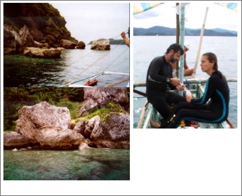
Figure 8. View of some reference
sampling sites at Gaspar Island, in the
marine reserve of Tres Reyes, and Drs.
Carr and Nipper processing porewater
samples on board an outrigger.
Materials and Methods
Sampling sites and collection methods
Pore water and/or sediment and
overlying water samples were collected
from 12 stations off Marinduque Island (Fig.
3) in October 2000. Sampling site
designation, details and coordinates are
presented in Table 1. Sediment samples and
water overlying the sediment surface were
collected at several stations (Table 1) for
chemical analyses. Pore water for toxicity
testing and chemical analyses was collected
in situ, by divers, at stations 1-6 and 10-12.
Weather and sea conditions did not permit in
situ sampling by diving at stations 7, 8 and
9. Therefore, sediment cores were collected
at knee depth at these three stations and pore
water was extracted on shore.
Sediment and overlying water were
collected directly into pre-cleaned 250 ml
high-density polyethylene containers with
screw caps and kept on ice for up to three
days, until arrival in Manila and shipping to
the USA. Pore water was extracted by
vacuum, using a device that consisted of a
ground glass aquarium air stone as the
filtration medium, attached to a 60-ml high
density polyethylene syringe (Fig. 9). All
sampling material was pre-soaked in
distilled water for a minimum of 24 hours
prior to use, and syringes were acid-washed.
Samples were removed from the syringes
and placed in pre-cleaned 500 ml high-

8
density polyethylene containers, and kept in
coolers on ice until arrival at the hotel.
Porewater samples were then filtered
through acid washed, binder free
borosilicate glass fiber filters with pore size
of 0.7 µm. Samples to be used for toxicity
testing were placed in pre-cleaned 250 ml
high density polyethylene jars with Teflon
lined screw caps, and an aliquot of the
filtered pore waters for chemical analyses
was placed in 50 ml high density
polyethylene containers. The filters used for
porewater filtration were of the kind
routinely used in chemistry laboratories for
the filtration of samples to be analyzed for
metals. These filters may have removed
other categories of contaminants from the
samples, if they were present, but since the
present survey was conducted to determine
environmental effects caused by metals
contamination from the mine tailings,
removal of other contaminants by the
filtration procedure was deemed acceptable.
Filtered samples were kept in coolers on ice
for up to 3 days, until arrival in Manila,
whereupon they were shipped to the USA by
Federal Express, accompanied by sample
tracking sheets. Water samples for toxicity
testing were shipped on dry ice, whereas
water and sediment samples for chemical
analyses were shipped on blue ice. Upon
arrival at the USGS toxicity testing
laboratory in Corpus Christi, Texas, the
samples were logged into the laboratory
sample tracking systems. The samples for
toxicity testing were placed in a freezer at -
20°C, and those for chemical analyses were
shipped on blue ice to the USGS chemistry
laboratory in Denver, Colorado.
Table 1. Description of sampling sites and related information
Site Site Latitude/Longitude Site Description Depth Type of sample
No. Designation (m) collected
Hakupan 13E33.08N/121E56.66 ~200 m S of Hakupan Island 15.0 Pore water (PW)
Island E
Calancan 13E33.70N/121E58.30 ~200 m W of tip of Calancan 2.0 PW, sediment (S),
Causeway E Causeway overlying water (OW)
Calancan 13E32.84N/121E57.58 ~200 m W of Calancan 7.5 PW
Cove E Causeway and ~400 m N of
mainland
Botilao 13E32.65N/121E56.20 ~300 m N of mainland and 9.0 PW
E directly S of Hakupan Island
Dinahunan 13E32.26N/121E55.62 ~300 m W of shore and ~500 6.5 PW
E NW of oil tank and pier
Guisian 13E32.83N/121E53.56 ~100 m from shore – 7.6 PW, S, OW
E believed to be “unimpacted”
area in aerial photo (Fig. 11
in Plumlee et al., 2000)
CMI 13E30.04N/121E50.66 ~200 m N of CMI causeway, 1.5 PW, S
Causeway E ~20m from shore
8 Mogpog River 13E29.05N/121E50.31 South side of river mouth 0.5 PW, S
E
9 Boac River 13E27.04N/121E48.70 North side of mouth of 0.2 PW
E northern tributary
10 Gaspar Island- 13E14.72N/121E51.82 South side of Gaspar Island 12.0 PW
south E ~50 m from shore
11 Gaspar Island- 13E14.95N/121E50.95 West side of Gaspar Island 12.0 PW, S, OW
west E ~50 m from shore
12 Gaspar Island- 13E15.20N/121E51.67 North side of Gaspar Island 12.0 PW
north E ~50 m from shore
1
2
3
4
5
6
7

9
Figure 9. Porewater sampling device
Porewater Quality Measurement and
Adjustment
Two days before conducting the toxicity
tests, the samples were moved from the
freezer to a refrigerator at 4°C. One day
prior to testing samples were thawed in a
tepid (20°C) water bath. Temperature of the
samples was maintained at 20 ± 1°C.
Sample salinity was measured and adjusted
to 30 ± 1
o
/
oo
using purified deionized water
or concentrated brine (see SOP F10.12,
Appendix 1). Other water quality
measurements (dissolved oxygen, pH,
sulfide, and ammonia concentration) were
made. Dissolved oxygen (DO) was
measured with an YSI
meter, salinity was
measured with a Reichert
refractometer,
and pH, and total ammonia (expressed as
nitrogen; NH
4
) were measured with an
Orion
meter and the respective probes.
Unionized ammonia (expressed as nitrogen;
NH
3
) concentrations were calculated for
each sample using the respective salinity,
temperature, pH, and NH
4
values.
Following water quality measurements and
adjustments, the samples were stored
overnight at 4°C but returned to 20 ± 1°C
before the start of the toxicity tests.
Toxicity Testing with Sea Urchin Gametes
and Embryos
Toxicity of the pore water was
determined using the fertilization and
embryological development tests with the
sea urchin Arbacia punctulata following the
procedures outlined in SOP F10.6 and F10.7
(Appendices 2 and 3).
Arbacia punctulata urchins used in this
study were obtained from Gulf Specimen
Company, Inc. (Panacea, Florida). A
standard reference porewater sample
collected from Redfish Bay, Texas, which
had been extracted by pneumatic pressure
and thereafter handled identically to the test
samples, was included with the toxicity tests
as a negative control, in addition to the
reference samples from Gaspar Island,
Philippines. The standard site for reference
porewater collection in Redfish Bay is far
removed from any known sources of
contamination and has been used
extensively as a reference site (Carr and
Chapman, 1992; National Biological
Survey, 1993, 1994; National Biological
Service 1995a, b; Carr et al., 1996a, b, 2000;
U.S. Geological Survey, 1997, 1998, 1999;
Nipper and Carr, 2001). In addition,
dilution blanks of filtered seawater and brine
controls (purified deionized water with brine
added to reach a 30
o
/
oo
salinity) were also
included. A dilution series test with a
reference toxicant, sodium dodecyl sulfate
(SDS), was included as a positive control.
Reference toxicant test results were
compared to a control chart with the results
of the previous tests with this reference
toxicant, in order to ensure that the
sensitivity of the test organisms was within
the standard range (Environment Canada,
1990).
Toxicity Test Data Analysis
The standard statistical analyses for
these toxicity tests involve comparisons
among treatments using ANOVA and
10
Dunnett's test. Prior to statistical analysis,
the transformed data sets were screened for
outliers (SAS Institute Inc., 1992) by
comparing the studentized residuals to a
critical value from a t-distribution chosen
using a Bonferroni-type adjustment. The
adjustment is based on the number of
observations, n, so that the overall
probability of a type I error is at most 5%.
The critical value, cv, is given by the
following equation: cv = t(df
Error
, .05/(2 x
n)). No outliers were detected. The
transformed data sets were tested for
normality and for homogeneity of variance
using SAS/LAB
Software (SAS Institute
Inc., 1992). Reference toxicant test results
were analyzed by the trimmed Spearman-
Karber method (Hamilton et al., 1977) with
Abbott's correction (Morgan, 1992) to
calculate EC
50
(effective concentration to
50% of the test organisms) values.
A second criterion was also applied to
compare test means to reference means.
Detectable significance criteria (DSC) were
developed to determine the 95% confidence
value based on power analysis of all similar
tests performed by our laboratory (Carr and
Biedenbach, 1999). This value is the
percent minimum difference from the
reference that is necessary to detect a
significant effect while minimizing type I
errors. The DSC value for the sea urchin
fertilization assay is 15.5% at _ #0.05, and
19% at _# 0.01. For the embryological
development test the DSC values at _
#0.05 and _# 0.01 are 16.4 and 20.6%,
respectively.
Chemical analyses of pore waters and
sediments
Pore waters were analyzed for major
cations and selected trace metals and
metalloids using inductively coupled
plasma-mass spectrometry. A detailed
description of the methods applied for the
chemical analyses and quality assurance
procedures is described in Lamothe et al.
(1999). It is believed that chloride
interference with the copper analyses caused
an increase of approximately 10 µg/L in the
measured concentrations. Therefore, this
was accounted for in the interpretation of
results. However, the copper values
measured at stations 8 and 9 are expected to
be accurate due to their low salinity (Table
2), thus lacking chloride interference.
Results and Discussion
Pore Water Quality Measurements
Water quality measurements for all
porewater samples, including the reference
pore water from Redfish Bay, Texas, as well
as the 0.45 µm filtered seawater control, are
presented on Table 2. Salinity was the most
variable of the measured porewater quality
parameters, ranging from 2 to 38 ppt. All
samples required salinity adjustment prior to
toxicity testing in order to satisfy the test
salinity requirement of 30 ± 1 ppt. The
percentage of pore water in the original test
samples (%OS) after salinity adjustment
(Table 2) ranged from 75 to 96%. Dissolved
oxygen was above 80% saturation in all
samples, and pH ranged from 7.43 and 8.25.
Unionized ammonia (NH
3
) was less than 5.4
µg/L in all samples from Marinduque. The
Redfish Bay reference pore water had a
somewhat higher pH (8.38), as well as
higher NH
3
concentration, of 57.8 µg/L,
which is still below toxic levels to A.
punctulata fertilization and embryological
development (Carr et al., 1996a).
Sea Urchin Toxicity Tests
Raw data and means from the
fertilization test are given in Table 3, and for
the embryological development test, in
Table 4. Only the two porewater samples
nearest to the Calancan mine tailings
causeway were significantly toxic to sea
urchin (A. punctulata) early-life stages. The

11
sample from station 2, closest to the
causeway, was the most toxic overall,
causing significant adverse effects in both
tests even at a dilution of 25%, whereas the
sample from station 3 was toxic in both tests
at the 50% dilution (see Fig. 10). The
gradient sampling performed in the area of
the Calancan causeway was in the
predominately up-current direction. Based
on the toxicity of the two samples collected
closest to the causeway, it is likely that
environmental effects caused by the tailings
would be more widespread in the down-
current direction. The EC
50
values for the
SDS tests (Tables 3 and 4) were within the
normal range based on the control chart with
the results of previous tests performed in our
laboratory with this reference toxicant,
indicating that the sensitivity of the test
organisms was within the acceptable
standard.
In previous surveys, pore water from
sandy sediment collected in the vicinity of
coral reefs in southeastern Mexico and in
Honolulu, Hawaii, exhibited toxicity,
showing that these kinds of tests are useful
indicators of contaminant accumulation in
toxic amounts around coral reefs, and could
serve as early warning signals of potential
biological effects to the reef itself (Nipper
and Carr, 2001). The results obtained in the
current study suggest that the area adjacent
to the Calancan causeway is adversely
affected by the mine tailings disposal.
Visual observations at the sites also
indicated biological degradation, with only
one species of small fish observed at station
2 and heavy siltation and smothered coral
seen at station 3 (see next section). Pore
water from stations 4 and 5 was not toxic to
sea urchin early life stages, but a large
amount of silt covering corals was still
observed at those sites, suggesting that
although there was no evidence of biological
degradation caused by chemical
contamination, a physical impact might be
caused by siltation due to erosion of fine
particulate matter from land.
Table 2. Water quality measurements of samples used for toxicity testing
Sample Original Adjusted OUS
1
DO
2
DO pH NH
4
3
NH
3
4
Sulfide
Salinity Salinity
(ppt) (ppt) (%) (mg/L) % sat (mg/L)
( g/L)
(mg/L)
Redfish Bay Ref. 40 30 75 6.69 90.7 8.38 0.803 57.8 <0.01
1 34.5 30 88 6.67 90.2 7.61 <0.1 <1.3 <0.01
2 34 30 88 6.93 93.1 7.52 <0.1 <1.1 <0.01
3 38 30 75 7.15 95.9 7.49 <0.1 <1.1 <0.01
4 26 30 96 6.79 91.9 7.72 <0.1 <1.7 <0.01
5 34 30 88 6.86 92.2 7.550 <0.1 <1.1 <0.01
6 35 30 86 6.87 92.3 7.43 <0.1 <0.9 <0.01
7 32 30 94 6.92 92.8 7.60 0.137 1.7 <0.01
8 2 30 75 7.00 94.00 8.25 <0.1 <5.4 <0.01
9 2 30 75 6.94 92.9 8.09 <0.1 <3.8 <0.01
10 35 30 87 6.75 91.0 8.05 <0.1 <3.5 <0.01
11 35 30 87 6.87 92.5 7.51 0.119 1.2 <0.01
12 35 30 86 6.77 90.9 7.49 <0.1 <1.0 <0.01
Brine control 0 30 75 6.36 87.8 8.41 <0.1 <7.7 <0.01
1
Percent of original undiluted sample after salinity adjustment;
2
Dissolved oxygen;
3
Total ammonia;
4
Unionized ammonia.

12
Figure 10. Map of Marinduque showing porewater toxicity data for sea urchin fertilization and
embryological development.
CMI
causeway
10
12
11
9
7
8
5
6
1
4
2
3
Tres Reyes
marine reserve
Calancan
tailings
causeway
Calancan
Bay
CMI
Pit
San Antonio Pit
Tapian Pit
Boac River
Makulapnit
River
Bol River
Mogpog River
0
150
Meters
N
Not toxic
<
0.01
Toxic at
>
25%,
<
0.01
Toxic at
>
50%,

13
Table 3. Mean (n=5) percent fertilization of the sea urchin, A. punctulata, in porewater samples
from Marinduque, reference and control samples, and in the reference toxicant test with SDS
Sample % Dilution Mean Fert. Standard EC
50
Significant
(%) Deviation (95% CI)
1
Effect
2
Redfish Bay 100 99.6 0.70
Redfish Bay 50 99.8 0.42
Redfish Bay 25 99.8 0.42
1 100 99.0 0.71
1 50 98.6 1.67 >100
1 25 98.6 0.55
2 100 0.8 1.30 **
2 50 0.2 0.45 <25 **
2 25 0.2 0.45 **
3 100 19.6 4.39 **
3 50 52.8 8.47 53.45 **
3 25 87.4 4.16 (46.99-60.80)
4 100 99.4 0.55
4 50 99.2 1.30 >100
4 25 99.4 0.89
5 100 100.0 0.00
5 50 100.0 0.00 >100
5 25 99.8 0.45
6 100 98.2 1.48
6 50 98.0 1.00 >100
6 25 97.2 2.49
7 100 96.8 1.48
7 50 97.2 1.79 >100
7 25 99.2 0.45
8 100 86.4 6.07
8 50 91.6 2.07 >100
8 25 93.0 3.54
9 100 100.0 0.00
9 50 99.4 0.89 >100
9 25 99.8 0.45
10 100 100.0 0.00
10 50 99.4 1.34 >100
10 25 100.0 0.00
11 100 98.8 2.68
11 50 99.2 0.45 >100
11 25 99.2 0.45
12 100 99.4 0.89
12 50 99.4 0.89 >100
12 25 99.2 0.84
Seawater Contr. 100 99.2 1.03
Brine control 100 96.6 0.55

14
Table 3. Continued
SDS (mg/L) 20 0.0 0.00
SDS (mg/L) 10 0.4 0.89
SDS (mg/L) 5 43.0 6.40 4.76
SDS (mg/L) 2.5 100.0 0.00 (4.45-5.10)
SDS (mg/L) 1.25 99.8 0.45
1
95% confidence interval in parenthesis;
2
Significantly different from the reference and below DSC at α≤0.01.
Table 4. Mean (n=5) percent normal embryological development of the sea urchin, A.
punctulata, in porewater samples from Marinduque, reference and control samples, and in the
reference toxicant test with SDS.
Sample % Dilution Mean Normal Standard EC
50
Significant
Larvae (%) Deviation (95% CI)
1
Effect
2
Redfish Bay 100 96.1 3.35
Redfish Bay 50 97.9 2.13
Redfish Bay 25 99.0 1.05
1 100 93.2 2.39
1 50 98.2 1.48 >100
1 25 98.6 0.89
2 100 0.0 0.00
**
2 50 0.0 0.00 <25
**
2 25 0.0 0.00
**
3 100 0.0 0.00
**
3 50 44.8 11.99 48.05
**
3 25 93.6 0.55 (44.60-51.76)
4 100 94.0 1.58
4 50 96.4 1.14 >100
4 25 97.6 1.52
5 100 96.4 1.34
5 50 96.2 2.17 >100
5 25 99.0 0.71
6 100 95.6 1.14
6 50 95.4 2.41 >100
6 25 96.4 2.61
7 100 94.4 2.30
7 50 96.6 2.30 >100
7 25 96.6 1.52
8 100 93.2 2.49
8 50 93.8 2.77 >100
8 25 98.2 0.84
9 100 96.8 1.64
9 50 96.2 1.48 >100
9 25 99.2 0.84

15
Table 4. Continued
10 100 96.2 2.77
10 50 95.6 2.30 >100
10 25 98.0 0.71
11 100 98.2 1.30
11 50 98.0 0.71 >100
11 25 98.2 1.64
12 100 94.6 2.07
12 50 96.2 1.48 >100
12 25 98.6 0.89
Seawater Contr. 100 97.7 2.41
SDS (mg/L) 20 0.0 0.00
SDS (mg/L) 10 0.0 0.00 3.46
SDS (mg/L) 5 0.2 0.45 (3.38-3.55)
SDS (mg/L) 2.5 93.6 3.21
SDS (mg/L) 1.25 98.0 1.87
1
95% confidence interval in parenthesis;
2
Significantly different from the reference and below DSC at α≤0.01
Visual observations of sampling stations
Station 1: Healthy appearance, with
small reef patches exhibiting small stag horn
coral, gorgonians, large basket sponges,
crinoids, starfish, and a variety of fish.
Station 2: Coarse sand. The only visible
life was one species of small burrowing fish.
Station 3: Heavy siltation, with large
amount of smothered hard coral. Sabellids,
crinoids and echinoids were observed, and
some small reef fish were seen in the area.
Station 4: Siltation was still observed,
but not as strongly as at station 3, although a
large amount of coral debris was present.
There were a large variety of hard corals,
crinoids, starfish, nudibranch mollusks and
reef fish.
Station 5: Heavy siltation and coral
suffocation with only a few reef fish seen.
Station 6: Healthier appearance, with
wide variety of corals, fish, and crinoids.
Large groups of one species of sea urchin
were observed, as well as starfish, large
bivalves, basket sponges, and sabellid
polychaetes.
Stations 7, 8 and 9: Sampling was
performed in shallow waters without diving,
for safety reasons, due to inclement weather.
Water was turbid and no visual observations
were made. The sediment was mostly
composed of coarse pebbles.
Stations 10, 11 and 12: These were the
reference stations at the marine reserve, off
Gaspar Island. They presented a healthy
appearance, with large biological diversity,
although little hard coral was present in the
area. There were many different species of
reef fish not seen at any other stations, giant
clams (Tridacna sp.) and nudibranch
mollusks, basket sponges, anemones and
gorgonians, tunicates, crustaceans, and a
variety of echinoderms, including
ophiuroids and sea urchins. Six different
species of sea urchins were observed at
station 12.
Chemical analyses
Chemical analyses of the porewater
samples exhibited elevated concentrations of
several metals at the toxic stations,
particularly Cd, Co, Cu, Ni, Pb and Zn at
station 2, and Co, Ni and Zn at station 3
(Table 5). In a comparison of the
concentrations of some selected metals with
the USEPA acute and chronic saltwater
criteria, it can be observed that criteria for

1
2
3
4
5
6
7
8
9
10
11
12
1
2
3
4
5
6
7
8
9
10
11
12
16
copper were exceeded at stations 2, 7, 8, and
9 in the vicinity of the CMI tailings
causeway and at the mouth of the Mogpog
river (Table 6). Acute and chronic criteria
for zinc were exceeded at stations 2 and 3,
and the chronic criteria for lead and nickel
were exceeded at station 2, which also had
the most toxic porewater sample.
Additional chemical and mineralogical
analyses of sediments collected at several
sites are still in progress and will be reported
in subsequent publications.
Table 5. Concentration of metals in pore waters of Marinduque Island.
Station Ag Al Ba Be Ca Cd Co Cr Cu
1
Fe K
(µg/L)
mg/L
(µg/L)
<5 450 30 <0.5 460 <0.2 <0.2 <10 10 890 460,000
<5 120 78 <0.5 480 2 5 <10 300 930 470,000
<5 56 62 <0.5 500 <0.2 3 <10 10 850 480,000
<5 43 52 <0.5 490 <0.2 <0.2 <10 10 770 480,000
<5 49 50 <0.5 490 <0.2 0.4 <10 10 710 480,000
<5 50 72 <0.5 480 <0.2 0.5 <10 10 660 470,000
<5 74 72 <0.5 450 <0.2 0.8 <10 20 620 410,000
<5 520 50 <0.5 13 <0.2 <0.2 <10 30 350 11,000
<5 170 40 <0.5 41 <0.2 <0.2 <10 8 140 11,000
<5 83 66 <0.5 470 <0.2 <0.2 <10 10 630 460,000
<5 75 57 <0.5 490 <0.2 0.4 <10 10 650 470,000
<5 230 40 <0.5 480 <0.2 0.4 <10 10 630 470,000
Table 5. Continued
Station Mg Mn Na Ni Pb Sb Tl V Zn
(mg/L)
(µg/L)
mg/L
(µg/L)
1,500 5.8 >3000 <1 <0.5 0.5 <0.5 2 20
1,500 54 >3000 10 18 0.4 <0.5 <2 320
1,500 17 >3000 6 0.7 0.4 <0.5 <2 100
1,400 17 >3000 <1 <0.5 0.5 <0.5 5 10
1,400 22 >3000 <1 <0.5 0.3 <0.5 4 20
1,400 14 >3000 1 <0.5 0.3 <0.5 3 20
1,200 160 >3000 <1 <0.5 <0.2 <0.5 <2 30
18 7 190 <1 1 <0.2 <0.5 <2 10
34 3.2 230 <1 <0.5 <0.2 <0.5 <2 9
1,400 7 >3000 <1 <0.5 0.2 <0.5 <2 20
1,400 13 >3000 1 <0.5 <0.2 <0.5 <2 10
1,400 17 >3000 <1 <0.5 0.2 <0.5 <2 20
1
Copper concentrations are likely to be 10 µg/L above true levels, due to chloride interference with the
measurements, except for stations 8 and 9, with 2 ppt salinity (see Table 2).

17
Table 6. U.S.EPA acute (short-term exposure) and chronic (long-term exposure) water quality criteria for selected
metals (in µg/L) in saltwater (USEPA, 1998), and station numbers at which criterion concentrations were exceeded
in the filtered samples. Criteria are not available for other metals analyzed.
Metal Acute Chronic Acute criteria exceedances Chronic criteria exceedances
Criteria Criteria Stations Stations
Cadmium 42 9.3
Zinc 90 81
2, 3 2, 3
Copper
1
4.8 3.1
2, 7, 8 2, 7, 8
Lead 210 8.1
2
Silver 1.9
NA
2
Nickel 74 8.2
2
1
10µg/L were subtracted from the measured values, to account for suspected chloride interference in the
measurements.
2
NA: Not Applicable. The method detection level was above the acute water quality criterion for silver.
Summary and Conclusions
High toxicity was observed and elevated
levels of metals were measured in the pore
waters collected at the two stations nearest
to the Calancan tailings causeway, although
those sites were in the predominately up-
current direction. Visual observations also
indicated biological degradation, with heavy
siltation and smothered coral at stations 2
through 5, suggesting that although samples
from stations 4 and 5 were not toxic to sea
urchin early life stages and did not present
particularly elevated levels of metals, a
physical impact might be caused by
siltation.
The toxicity data and the visual
observations suggest that the re-
establishment of the benthic community, as
well as of coral reefs and their associated
fauna and flora, is unlikely to occur in the
vicinity of the Calancan causeway. The
elevated concentration of copper in the pore
water from the station 2, closest to the
causeway, suggests that this causeway is a
constant source of copper contamination to
the surrounding environment and is
impairing the re-colonization of the area by
the local natural biota.
This preliminary survey suggests that
effects related to past mining activities are
still evident and warrants a more
comprehensive study to assess the severity
and areal extent both up and down current of
the tailings causeways.
Acknowledgements
We thank Congressman Edmund O.
Reyes for the invitation to conduct this
survey on Marinduque, and we are grateful
for the warm welcome and continued
logistical assistance given to us by the staff
of Governor Carmencita Reyes during our
stay in Marinduque. We are indebted to the
expert technical assistance of Jim
Biedenbach, Russell Hooten, and Linda May
who processed the samples in the laboratory
and conducted the toxicity tests, and to Al
Meier and Peter Theodorakos for
performing the chemical analyses. We
thank Drs. Jack Medlin and Tom May for
their helpful reviews of this report.

18
Literature Cited
Carr, R.S. and J. M. Biedenbach. 1999. Use of power analysis to develop detectable significance
criteria for sea urchin toxicity tests. Aquat. Ecosys. Health Manage. 2:413-418.
Carr, R.S. and D.C. Chapman. 1992. Comparison of solid-phase and pore-water approaches for
assessing the quality of marine and estuarine sediments. Chem. Ecol. 7:19-30.
Carr, R.S., D.C. Chapman, C.L. Howard, J.M. Biedenbach. 1996a. Sediment quality triad
assessment survey of the Galveston Bay, Texas system. Ecotoxicology 5:341-364.
Carr, R.S., E.R. Long, H.L. Windom, D.C. Chapman, G. Thursby, G.M. Sloane, D.A. Wolfe.
1996b. Sediment quality assessment studies of Tampa Bay, Florida. Environ. Toxicol. Chem.
15:1218-1231.
Carr, R.S., P.A. Montagna, J.M. Biedenbach, R. Kalke, M.C. Kennicutt, R. Hooten, G. Cripe.
2000. Impact of storm-water outfalls on sediment quality in Corpus Christi Bay, Texas,
USA. Environ. Toxicol. Chem. 19:561-574.
Environment Canada. 1990. Guidance document on control of toxicity test precision using
reference toxicants. Environmental Protection Series, Report EPS 1/RM/12. Environment
Canada, Ottawa, Ontario, Canada.
Hamilton, M.A., R.C. Russo, R.V. Thurston. 1977. Trimmed Spearman-Karber method for
estimating median lethal concentrations in toxicity bioassays. Environ. Sci. Technol. 11:714-
719; Correction 12:417 (1978).
Lamothe, P.J., A.L. Meier, S. Wilson. 1999. The Determination of Forty Four Elements in
Aqueous Samples by Inductively Coupled Plasma - Mass Spectrometry. U.S. Geological
Survey, Open-File Report 99-151, Denver, CO, 14 p.
Morgan, B.J.T. 1992. Analysis of Quantal Response Data, London, England: Chapman and Hall,
511 pp.
National Biological Service (NBS). 1995a. Toxicity testing of sediments from Biscayne Bay and
Surrounding Areas. Report submitted by the National Biological Service to the National
Oceanic and Atmospheric Administration, Coastal Monitoring and Bioeffects Division,
Seattle, WA, 11 pp. + 17 tables, 11 figures and 4 attachments.
National Biological Service (NBS). 1995b. Toxicity testing of sediments from western Florida
and coastal South Carolina and Georgia. Report submitted by the National Biological
Service to the National Oceanic and Atmospheric Administration, Coastal Monitoring and
Bioeffects Division, Seattle, WA, 14 pp. + 35 tables, 10 figures and 4 attachments.
National Biological Survey (NBS). 1993. Toxicity testing of sediments from Charleston Harbor,
South Carolina and vicinity. Report submitted by the National Biological Survey to the
National Oceanic and Atmospheric Administration, Ocean Assessment Division, Seattle,
WA, 7 pp. + 16 tables and 4 attachments.
National Biological Survey (NBS). 1994. Survey of sediment toxicity in Pensacola Bay and St.
Andrew Bay, Florida. Report submitted by the National Biological Survey to the National
Oceanic and Atmospheric Administration, Ocean Assessment Division, Seattle, WA, 12 pp.
+ 24 tables and 5 attachments.
Nipper, M. and R.S. Carr. 2001. Porewater toxicity testing: a novel approach for assessing
contaminant impacts in the vicinity of coral reefs. Bull Mar Sci 68 (in press).

19
Plumlee, G.S., R.A. Morton, T.P. Boyle, J.H. Medlin, J. Ceteno. 2000. An Overview of Mining-
related Environmental Issues, Marinduque Island, Philippines: Observations from a Joint
U.S. Geological Survey – Armed Forces Institute of Pathology Reconnaissance Field
Evaluation, May 12-19, 2000. Denver, Colorado: U.S. Geological Survey, Open-File Report
00-397. 36 p.
http://geology.cr.usgs.gov/pub/open-file-reports/ofr-00-0397/
SAS Institute Inc. 1992. SAS/LAB Software: User's Guide, Version 6, First Edition, Cary, NC:
SAS Institute Inc., 291 pp.
U.S. Environmental Protection Agency (USEPA). 1998. National recommended water quality
criteria; republication. Federal Register, Part IV, v. 63, p. 68354-68364.
U.S. Fish and Wildlife Service (USFSW). 1992. Amphipod solid-phase and sea urchin porewater
toxicity tests of Tampa Bay, Florida sediments. Report submitted by the U.S. Fish and
Wildlife Service to National Oceanic and Atmospheric Administration, Ocean Assessment
Division, Seattle, WA, 9 pp. + 19 appendices.
U. S. Geological Survey (USGS). 1997. Toxicity testing of sediments from Biscayne Bay,
Florida and surrounding areas - Phase II. Report submitted by the USGS to National
Oceanic and Atmospheric Administration, Coastal Monitoring and Biological Assessment
Division, Seattle, WA, 10 pp. + 8 tables, 10 figures and 4 attachments.
Sampling team left to right: Joven Lilles, Dr. Scott Carr, Dindo Asuncion, unidentified
driver, driver Dan, Ton Monteagudo, Erwin Penafiel, and Dr. Marion Nipper (the photographer).
APPENDIX 1
SOP F10.12
WATER QUALITY ADJUSTMENT OF SAMPLES
Corpus Christi SOP: F10.12 Page 1 of 6
Date Prepared: March 14, 1991
Date Revised: May 17, 1994
WATER QUALITY ADJUSTMENT OF SAMPLES
1.0 OBJECTIVE
In order to perform toxicity tests with saline samples, all test and reference samples should
be similar in salinity so that salinity is not a factor in survival of test organisms.
Additionally, dissolved oxygen (DO) concentrations should be sufficiently high to ensure
that low DO is not a source of stress to the test organisms. At the Corpus Christi field
station, toxicity tests are performed using a variety of marine and estuarine organisms,
including the sea urchin Arbacia punctulata, the polychaete Dinophilus gyrociliatus, the
harpacticoid copepod Longipedia sp., and the red drum Sciaenops ocellatus. The aqueous
samples tested may be pore water, different kinds of discharges and effluents, surface
microlayer, or subsurface water samples that may range in salinity from 0-36
o
/
oo
.
Although from test to test salinities used in the different toxicity tests may vary, the
individual toxicity tests performed on a particular day are run at a single target salinity.
Since initial salinities of the porewater or water samples to be tested commonly vary, they
will require salinity adjustment to within 1
o
/
oo
of the target salinity. Additionally, DO
should normally be 80% saturation in all samples tested.
2.0 PREPARATION
2.1 Equipment and Labware
The supplies and equipment needed are listed in Attachment 1.
2.2 Source of Dilution Water
For samples lower in salinity than target salinity, concentrated brine (_100
o
/
oo
) is
added to increase salinity. Concentrated brine is prepared by heating (to 35-40°C)
and gently aerating filtered natural seawater (1 µm) to concentrate the salts by
evaporation. For samples higher in salinity than target salinity, HPLC ultrapure
sterile water (J.T. Baker® Cat. #JT4218-2) is added to decrease salinity.
3.0 PROCEDURES
The following describes the procedures required for the adjustment and determination
of specific water quality parameters of a sample.
Corpus Christi SOP: F10.12 Page 2 of 6
3.1 Preparation for Salinity Adjustment
1. Although fresh samples are routinely tested at the Corpus Christi field station,
most of the samples tested are stored frozen in amber I-Chem® jars. If frozen,
remove samples from freezer and allow them to thaw at room temperature or
immerse them in a tepid water bath to thaw, ensuring that sample temperature does
not exceed 25°C. The samples may be thawed the day of water quality adjustment
(WQA) or may be transferred from the freezer to a refrigerator (4°C) the day
before WQA and then completely thawed the following day. After thawing, allow
the samples to come to room temperature. Generally, the samples should be
maintained at the same temperature required for the toxicity test that will be
conducted. The temperature requirement for most toxicity tests performed at this
field station is 20±1°C, and room temperature should be maintained accordingly.
2. Turn bottled sample end over end a few times to mix thoroughly before measuring
salinity. Using a salinity refractometer, measure salinity and record on Water
Quality Adjustment Data Form (Attachment 2).
3. In order to make calculations for the salinity adjustment, the volume of the sample
must be known. When porewater or other water samples are collected and
transferred to amber jars for storage, they are commonly measured to an
approximate volume (110 mL, for example) prior to freezing. On the day of
WQA, this volume should be recorded on the WQA data form for the respective
samples. If the volume is unknown at this point, it should be measured using a
graduated cylinder of appropriate size, and recorded on the data sheet.
3.2 Salinity Adjustment
3.21 Reducing the salinity of aqueous samples
Refer to the formulas below to calculate the volume of HPLC water needed to
reduce the initial sample salinity to the target salinity. Add the volume calculated,
mix the bottle thoroughly, check the salinity with a refractometer, and record the
volume of HPLC water added as well as the final salinity.
(i) (target
o
/
oo
÷ sample
o
/
oo
) _ sample vol. in mL = A
(ii) sample vol ÷ A = B
(iii) sample vol. ÷ A = C
(iv) B _ C = volume of HPLC water to add
3.22 Increasing the salinity of aqueous samples
Refer to the formula below to calculate the volume of concentrated brine needed
to increase the initial sample salinity to the target salinity. Add the volume
calculated, mix the bottle thoroughly, check the salinity with a refractometer,
and record the volume of brine added as well as the final salinity.
Corpus Christi SOP: F10.12 Page 3 of 6
(i) ((target
o
/
oo
_ sample
o
/
oo
) _ sample vol. in mL) ÷ (brine
o
/
oo
_ target
o
/
oo
) =
vol. of brine to add
3.3 Dissolved Oxygen Adjustment
Measure and record DO and percent DO saturation of sample (SOP F10.13).
Occasionally, a sample will have DO of less than 80% saturation. Any such samples
should be gently stirred on a magnetic stirrer to increase the DO level above 80%.
Record initial DO, the elapsed mixing time, and final DO in the comments section of
the Water Quality Adjustment Data Form. (On the following day, DO should be
rechecked and brought to >80% by stirring again if necessary before the toxicity test
is performed.)
3.4 Other Water Quality Determinations
1. Measure pH (SOP F10.21) and record on the Water Quality Adjustment Data
Form.
2. Measure and record ammonia concentration (SOP F10.4).
3. Measure and record sulfide concentration if required.
4.0 DATA COLLECTION
All raw data are entered on one standardized form, the Water Quality Adjustment Data
Form (see Attachment 2) at the time the determinations or adjustments are made.
5.0 QUALITY CONTROL
A data form (Attachment 2) will be used to document all sample handling procedures for
each sample. The person(s) recording data on the sheet will initial each sheet. Original
data forms after completion will be stored in a three-ring file in the possession of the field
station leader. Copies will be kept in the lab.
6.0 TRAINING
Personnel who will perform this task should first read this protocol and then operate under
supervision during the preparation of at least two samples.

Corpus Christi SOP: F10.12 Page 4 of 6
7.0 SAFETY
The NaOH solution used in the ammonia determination procedure is a highly caustic
liquid. Care should be taken to avoid its contact with skin or clothing. Should such
contact occur, quickly flush affected with water. A sink is present along the west wall of
the dry lab, another is present along the east wall of the wet lab, and an eye flushing station
is present in the northwest corner of the wet lab near the entrance door. The samples
handled may be pore water, effluent, discharges, or other water samples that may contain
contaminants. Care should be taken to avoid contact with the samples.
8.0 ATTACHMENTS
Attachment 1. Equipment List for Water Quality Adjustment
Attachment 2. Water Quality Adjustment Data Form
Prepared by:
Duane C. Chapman
Fishery Biologist
Approved by:
R. Scott Carr
Field Station Leader
Anne E. Kinsinger
Chief, Field Research Division
Joseph B. Hunn
Quality Assurance Officer

Corpus Christi SOP: F10.12 Page 5 of 6
ATTACHMENT 1
EQUIPMENT LIST FOR WATER QUALITY ADJUSTMENT
Graduated cylinders
Pipetters
Latex gloves
Magnetic stirrer and stir bars
10 M NaOH
Concentrated brine (See section 2.2 for preparation)
HPLC ultrapure sterile water (J.T. Baker® #JT4218-2)
Salinity refractometer
Dissolved oxygen meter
pH electrode, buffer solutions, and meter
Ammonia electrode, standard solutions, and meter
Sulfide electrode, standard solutions, and meter
Data sheets
Hand calculator

Corpus Christi SOP: F10.12 Page 6 of 6
ATTACHMENT 2
WATER QUALITY ADJUSTMENT DATA FORM
STUDY PROTOCOL INITIALS _________
SAMPLE DESIGNATION DATE
A. Salinity Adjustment:
Initial volume (mL)
Initial salinity (
o
/
oo
)
Vol. Baker® HPLC water added (mL)
Vol.
o
/
oo
brine added (mL)
% of original sample
(initial vol./final vol. x 100)
B. Character of Sample (after salinity adjustment):
Final Volume (mL)
Final Salinity (
o
/
oo
)
pH
Dissolved oxygen (mg/L)
DO saturation (%)
Total ammonia (mg/L)
Sulfide (mg/L)
COMMENTS
APPENDIX 2
SOP F10.6
SEA URCHIN FERTILIZATION TOXICITY TEST
Corpus Christi SOP: F10.6 Page 1 of 15 pages
Date Prepared : April 10, 1990
Date Revised: March 10, 1995
SEA URCHIN FERTILIZATION TOXICITY TEST
1.0 OBJECTIVE
The purpose of the fertilization toxicity test with the sea urchin, Arbacia punctulata, is to
determine if a sea water, pore water, sea surface microlayer, or other sample reduces
fertilization of exposed gametes relative to that of gametes exposed to a reference sample.
The test may also be used to determine the concentration of a test substance which reduces
fertilization. Test results are reported as treatment (or concentration) which produces
statistically significant reduced fertilization or as concentration of test substance which
reduces fertilization by 50 percent (EC
50
). This test can be performed concurrently with Sea
Urchin Embryological Development Toxicity Test (SOP 10.7) and/or Sea Urchin
Genotoxicity/Teratogenicity Test (SOP 10.8), using the same pretest and sperm and egg
collection.
2.0 TEST PREPARATION
2.1 Test Animals
Gametes from the sea urchin, Arbacia punctulata are used in the sea urchin fertilization
toxicity test. Animals can be collected in the field or obtained from a commercial supplier.
A. punctulata can be differentiated from other species of urchins which are found in Texas
by the five plates surrounding the anal opening, and by round sharp spines on the dorsal
surface of the test and flattened spines surrounding the Aristotle's lantern. Urchins can be
maintained easily in aquaria or other tanks with running seawater or an aquarium filter.
Urchins will eat a wide variety of marine vegetation. A good diet may be provided by
placing rocks from jetties (which have been colonized by diatoms and macroalgae) into the
tank with the urchins or romaine lettuce may be provided as a substitute. Temperature
manipulations of the cultures will prolong the useful life of the urchins. Cultures are
maintained at 16 ± 1EC when gametes are not required. Temperature is gradually increased
to 19 ± 1EC at least one week prior to gamete collection and subsequently decreased if no
further tests are planned. Photoperiod is maintained at 16 hours of light per day. Water
quality parameters should be monitored weekly and salinity maintained at 30 ± 3
o
/
oo
. Males
and females should be kept in separate tanks.

Corpus Christi SOP: F10.6 Page 2 of 15 pages
2.2 Dilution Water
HPLC reagent grade purified water or concentrated seawater brine is used to adjust samples
to 30
o
/
oo
as described in Water Quality Adjustment of Samples (SOP 10.12). Concentrated
seawater brine (90-110
o
/
oo
) is made in large batches by heating seawater to 40°C or less in
large tanks with aeration for 3-4 weeks. Brine quality will remain constant over long periods
with no refrigeration. At the time of salinity adjustment, pH, ammonia, and dissolved
oxygen are also measured. Salinity adjustment and water quality data are recorded on
prepared data forms.
Filtered (0.45 µm) seawater adjusted to 30
o
/
oo
is used to wash eggs and is also used for
sperm and egg dilutions. The acronym MFS (for Millipore® filtered seawater) is used for
this filtered and salinity adjusted seawater.
2.3 Test System: Equipment
When testing samples for potential toxicity, five replicates per treatment are recommended.
One replicate is a 5 mL volume of sample in a disposable glass scintillation vial. When
conducting a dilution series test, fifty percent serial dilutions may be made in the test vials,
using MFS as the diluent.
2.3.1 Equipment
A list of equipment necessary for conducting this test is given in Attachment 1
(Equipment List for Fertilization Toxicity Test).
2.3.2 Solutions
10% Buffered Formalin:
1,620 mL sea water
620 mL formaldehyde
6.48 g NaH
2
P0
4
or KH
2
PO
4
(mono)
10.5 g Na
2
HPO
4
or K
2
HPO
4
(dibasic)
1 mL needed for each replicate. Fill the dispenser.
2.4 Collection and Preparation of Gametes
Quality gametes must first be collected, and then diluted to the appropriate concentration
for addition to the test vial.
Corpus Christi SOP: F10.6 Page 3 of 15 pages
2.4.1 Selection of Urchins to be Used in Toxicity Test.
1. Take two or three females and place in shallow bowl, barely covering tests with
seawater.
2. Stimulate release of eggs from gonopores of a female by touching test with electrodes
from a 12V transformer.
3. Collect a few eggs from between spines using a 10 mL disposable syringe with a large
gauge blunt-tipped needle attached. Discard the first small quantity of eggs expelled
from each gonopore and continue collecting. Place a 2 to 5 drops of eggs onto a
scintillation vial containing 10ml of filtered seawater. Rinse syringe and repeat for
each female.
4. Select females which have round, well developed eggs, and which do not release
clumps of eggs or undeveloped ovarian tissue.
5. Place 2-4 males in shallow bowl(s) with a small amount of seawater, leaving the upper
1
/
2
to
1
/
3
of the animals uncovered.
6. Stimulate release of sperm from gonopores by touching test with electrodes from 12V
transformer (about 30 seconds each time). If sperm is watery, reject the animal and
choose another. Sperm should be the consistency of condensed milk. Collect sperm
using a pasteure pipette with a rubber bulb attached.
Generally, a gamete check is performed in order to ensure that both the male and the
female urchins used in the test have gametes with a high degree of viability. If the gamete check
is performed, two to five females (depending on confidence in the proportion of urchins in the
holding facility in good reproductive status) and at least two males should be selected using the
above procedures. The check is performed by adding 5 to 7 drops of a concentrated dilution of
sperm to the eggs in the scintillation vials (collected as described above) and observing the eggs
under the microscope after 10 minutes. The concentrated dilution of sperm is usually made by
diluting 20-50µl of sperm in 10 ml of filtered seawater. If the proportion of eggs fertilized is
high (95-100%), that female and male may be used in the pretest and test. Sperm from a number
of males or females may be combined in the beginning if the gamete check reveals a number of
high quality animals or the confidence is high in the quality of the gametes. Once a good male
and female are selected a pretest can be conducted to determine the correct dilution of sperm to
use in the test (Attachment 2).
2.4.2 Obtain Eggs
1. Place selected female in large Carolina dish and add enough water to cover the urchin's
test with approximately 1 cm of seawater. Stimulate release of eggs from female with
12V transformer.
Corpus Christi SOP: F10.6 Page 4 of 15 pages
2. Collect eggs as above using the 10 mL syringe. Remove needle before dispensing eggs
into a disposable shell vial or other clean container capable of holding 25-50 mL.
Collect enough eggs for pretest and test. If female stops giving eggs readily or starts
giving chunky material, cease stimulation and collection of eggs from that female.
3. Add MFS to fill shell vials, gently mixing eggs. Allow eggs to settle to bottom of vial.
Remove water with a pipette. Replace water, again gently mixing the eggs.
4. Repeat washing procedure.
2.4.3 Prepare Appropriate Egg Concentration
1. Put approximately 100 mL of 30
o
/
oo
MFS in a 250 mL beaker, and add enough
washed eggs to bring the egg density to approximately 10,000 per mL. If more than
400 total replicates (27 treatments) are to be tested, a larger amount of water and a
correspondingly larger amount of eggs should be used. Two hundred µL of this egg
solution will be used per replicate, and it is easier to maintain proper mixing and
uniform egg density if there is an excess of at least 50%.
2. Check egg density and adjust to within approximately 9000 to 11,000 eggs per mL, as
follows. Gently swirl egg solution until evenly mixed. Using a pipette, add 1 mL of
the solution to a vial containing nine mL seawater. Mix and transfer 1 mL of this
diluted solution to a second vial containing 4 mL of seawater. Again, mix and transfer
1 mL of this diluted solution to a counting slide such as a Sedgewick-Rafter slide.
3. Using a microscope (either a compound microscope with a 10x objective or a
dissecting scope may be used here), count the number of eggs on the slide. If the
number is not between 180 and 220, then adjust by adding eggs or water. If egg count
is > 220 use the following formula to calculate the amount of water to add:
("egg count" - 200/200) x Current Volume of Eggs = Volume seawater to add
to stock (mLs)
If egg count < 200 add a small amount of eggs. Since it is less arbitrary and more
likely to arrive at an acceptable count when using the water addition formula, it is
better to originally overestimate the amount of eggs to add to the 100 mL of water.
4. Repeat steps 2 and 3 until an acceptable egg count (between 180 and 220) is obtained.
2.4.4 Obtain Sperm
Place selected male urchin in a large Carolina dish containing 1-2 cm of water. About
half of test should be above water level. Stimulate male with 12V transformer, and
collect about 0.5 mL of unwetted sperm from between spines using a pasteur pipette.
Place sperm into a plastic microcentrifuge tube. Keep on ice until used. Be careful not to
Corpus Christi SOP: F10.6 Page 5 of 15 pages
add any water or sperm which has contacted water to the vials. High quality sperm
collected dry and kept on ice will last at least eight hours without measurable decline in
viability.
2.4.5 Prepare Appropriate Sperm Dilution
It is desirable for control fertilization to be within 60-90%. Although controls outside
these bounds do not automatically disqualify a test, particularly if a valuable dose
response is generated, the sensitivity of the test is reduced by fertilization rates greater
than 90% and good dose responses may be difficult to obtain with less than 60%
fertilization in controls. Density of sperm in the sperm solution should be determined
with this goal in mind. Condition of the animals and length of acclimation to the
aquarium may affect the chosen sperm density. The pretest (Attachment 2) may be used
to calculate an appropriate sperm dilution. Generally, a dilution of between 1:10,000 and
1:2500 will result in desirable fertilization rates, if the animals are in good condition.
For example, if a sperm dilution of 1:5000 is required (as determined from the pretest),
add 20 µL sperm to 10 mL MFS. Mix thoroughly, then add 1 mL of this solution to 9 mL
MFS. Sperm should not be wetted until just before starting the test. Sperm wetted more
than 30 minutes before the test has begun, including sperm dilutions used in any pretest,
should be discarded and a new dilution made from sperm kept on ice.
3.0 TEST PROCEDURES
1. Add 50 µL appropriately diluted sperm to each vial. Record time of sperm addition.
Sperm should be used within 30 minutes of wetting.
2. Incubate all test vials at 20 ± 2°C for 30 minutes. At this point it is useful to set a timer
for five to ten minutes prior to the end of the incubation period. This will notify the
worker early enough to be ready to start the next step exactly on time.
3. While gently swirling the egg solution to maintain even mixing of eggs, use a 200 µL
pipetter to add 200 µL diluted egg suspension to each vial. Pipette tips are cut back using
a clean razor blade to prevent crushing the eggs during pipetting. Record time of egg
addition.
4. Incubate for 30 minutes at 20 ± 2°C. The timer may be used again at this point.
5. Using the dispenser, add 1 mL of 10% buffered formalin to each sample.
6. Vials may now be capped and stored overnight or for several days until evaluated.
Fertilization membranes are easiest to see while eggs are fairly fresh, so evaluation within
two to three days may decrease the time required for evaluation.

Corpus Christi SOP: F10.6 Page 6 of 15 pages
7. If it is not possible to make the evaluations within several days or the membranes are
difficult to discern, an optional technique may be employed. Make up a 200
o
/
oo
NaCl
solution (pickling salt) and add 2 to 4 drops of the solution to a 1 mL egg sample on a
microscope slide. This solution causes the egg, but not the membrane, to shrink briefly
thereby making the membrane easier to see. The effect only lasts for a short time (~5
min.) so the observations must be made immediately after the NaCl solution is added. If
this optional technique is employed, it must be used on all samples in that test series.
4.0 DATA COLLECTION AND TABULATION
1. Transfer approximately 1 mL eggs and water from bottom of test vials to counting slide.
Observe eggs using compound microscope under 100X magnification. Dark field
viewing is useful here in identifying fertilization membranes.
2. Count 100 eggs/sample using hand counter with multiple keys (such as a blood cell
counter), using one key to indicate fertilized eggs and another to indicate unfertilized
eggs. Fertilization is defined by the presence of fertilization membrane surrounding egg.
3. Calculate fertilization percentage for each replicate test:
Total No. Eggs - No. Eggs Unfertilized x 100 = Percent Eggs Fertilized
Total No. Eggs
5.0 DATA ANALYSIS
Data are recorded on standardized data sheets (See Attachments 3-7). Normally, percent
fertilization in each treatment is compared to an appropriate reference treatment (seawater,
pore water or sea surface microlayer from an uncontaminated environment). Statistical
comparisons are made using analysis of variance (ANOVA) and Dunnett's t-test (Sokal and
Rohlf 1981) on the arc sine square root transformed data. For multiple comparisons among
treatments, Ryan's Q test (Day and Quinn 1989) with the arc sine square root transformed
data is recommended. The trimmed Spearman-Karber method with Abbott's correction is
recommended to calculate EC
50
values for dilution series tests (Hamilton et al. 1977)
6.0 QUALITY CONTROL
Quality control tests may be run using both positive and negative controls with multiple
replicates (as many as desired). Typically, a reference toxicant dilution series (sodium
dodecyl sulfate) is tested with each test to evaluate the effectiveness of the sperm dilution
chosen. Negative controls may include a reference porewater, filtered seawater, and/or a
reconstituted brine.
Corpus Christi SOP: F10.6 Page 7 of 15 pages
7.0 TRAINING
A trainee will conduct the test with supervision initially. Determining egg concentrations
and fertilization counts are test specific activities. These functions can be performed
independently after a trainee has demonstrated he or she can accurately reproduce the test.
8.0 SAFETY
The sea urchin fertilization toxicity test poses little risk to those performing it. Care should
be taken when making and dispensing the 10% buffered formalin solution; use a hood if
available, but make sure the test area is well ventilated. Protective gloves can be worn when
pipetting or dispensing formalin or potentially toxic samples.
Care should be taken when collecting or otherwise handling sea urchins. Urchin spines are
sharp and fragile and may puncture the skin and break off if handled roughly. First aid
similar to treatment of wood splinters is effective in this case (removal of spine and
treatment with antiseptic). Collection of sea urchins by snorkeling should not be done alone.
9.0 ATTACHMENTS
Attachment l. Equipment List for Fertilization Toxicity Test
Attachment 2. Pretest to Insure Selection of Quality Gametes
Attachment 3. Water Quality Adjustment Data Form
Attachment 4. Sea Urchin Pretest Data Sheet
Attachment 5. Sea Urchin Pretest Continuation Data Sheet
Attachment 6. Sea Urchin Fertilization/Embryological Development Toxicity Test Gamete
Data Sheet
Attachment 7. Sea Urchin Fertilization Toxicity Test Fertilization Data Sheet
10.0 REFERENCES
Day, R.W. and G.P. Quinn. 1989. Comparisons of treatments after an analysis of variance
in ecology. Ecol. Monogr. 59:433-463.
Hamilton, M.A., R.C. Russo, and R.V. Thurston. 1977. Trimmed Spearman-Karber method
for estimating median lethal concentrations in toxicity bioassays. Environ. Sci. Technol.
11(7):714-719; Correction 12(4):417 (1978)
Sokal, R.R., and F.J. Rohlf. 1981. Biometry. 2
nd
edition. W.H. Freeman and Company, San
Francisco, CA 859 pp.

Corpus Christi SOP: F10.6 Page 8 of 15 pages
Prepared by:
Duane Chapman
Fishery Biologist
Approved by:
R. Scott Carr
Field Station Leader
Anne E. Kinsinger
Chief, Field Research Division
Joseph B. Hunn
Quality Assurance Officer
Corpus Christi SOP: F10.6 Page 9 of 15 pages
Attachment 1
EQUIPMENT LIST FOR FERTILIZATION TOXICITY TEST
Large Carolina dishes (at least 2)
20 mL KIMBLE scintillation vials (These should be type shipped with caps off, and without cap
liners. If other brand or type is used, the vials should be tested for toxicity prior to use.)
400 mL beaker or wide-mouthed thermos for holding vials of sperm
250 mL beakers (4)
Pasteur pipettes and latex bulbs
plastic microcentrifuge tubes
25 mL shell vials or equivalent
Test tube rack (to hold shell vials)
12V transformer with pencil type electrodes
Styrofoam (or something to hold electrode tips)
10 cc syringe with large diameter blunt ended needle (make by grinding sharp point off the
needle with a grinding stone)
Marking pens
Ice
10-100 µL pipetter
50-200 µL pipetter
5 mL pipetters (2)
Counting slide such as Sedgewick-Rafter chamber
Compound microscope with 10x objective and dark field capability
Hand tally counter
Calculator
Timer for exposure / incubation periods
Buffered formalin and dispenser
Filtered (0.45 µm) seawater, adjusted to 30
o
/
oo
Data sheets
Baker reagent grade water
Approximately 100
o
/
oo
concentrated brine
Corpus Christi SOP: F10.6 Page 10 of 15 pages
Attachment 2
PRETEST TO INSURE SELECTION OF QUALITY GAMETES
1. Using the procedure in section 2.4.1, select 2 to 5 females and at least 2 male urchins to
be used in the pretest.
2. Fill pretest vials with five mL of reference water. There should be at least two vials for
each combination of male, female, and pretest sperm concentration (step 4 below). For
example, in a pretest with two females, one male, and six pretest sperm concentrations, 24
vials (2 X 2 X 6) would be needed. Arrange and mark vials accordingly in a rack.
3. Perform steps 2.4.2 (egg collection) and 2.4.3 (egg dilution) for each female urchin.
Make enough volume of the egg suspension to perform the pretest and the test.
4. Perform step 2.4.4 (sperm collection) for each male urchin or male combination. Prepare
a dilution series of sperm concentrations which will bracket the 60-90% fertilization rate in
the test. Sperm dilution will depend on the health and reproductive status of the male urchin,
but in most cases the following "standard dilution" should be used:
1: 250 (20 µL dry sperm added to 5 mL MFS. This concentration is used only as
stock solution to make up the rest of the dilution series and is not used full strength
in the pretest.)
1: 1250 (1 mL of 1:250 and 4 mL MFS)
1: 2500 (1 mL of 1:250 and 9 mL MFS)
1: 5000 (2 mL of 1:2500 and 2 mL MFS)
1: 7500 (2 mL of 1:2500 and 4 mL MFS)
1:10000 (3 mL of 1:7500 and 1 mL MFS)
1:12500 (1 mL of 1:2500 and 4 mL MFS)
Sperm must be used within 30 minutes of dilution. Leave undiluted sperm on ice and
retain, because a new sperm dilution of the concentration determined in this pretest will be
needed for the toxicity test. Sperm diluted for use in the pretest may not be used in the
toxicity test, because the time elapsed since the addition of water is too great.
5. As in section 3.0 add 50 µL of the diluted sperm to each pretest vial. Incubate for 30
minutes at approximately 20°C, and add 200 µL of the egg suspension. Incubate for another
30 minutes, then fix with 1 mL of the buffered formalin solution.
6. As in section 4.0, obtain a fertilization rate for the vials. There is no need to count all
vials, enough vials should be counted to determine a good male/female combination, and an
appropriate sperm dilution factor. If more than one male/female combination is acceptable,
this is a good opportunity to choose a female which exhibits easily visible fertilization
membranes or in cases where there are many samples, to combine eggs from different
females . The appearance of the fertilization membranes may vary among female urchins,
and presence of easily visible membranes facilitates counting.

Corpus Christi SOP: F10.6 Page 11 of 15 pages
Attachment 3
WATER QUALITY ADJUSTMENT DATA FORM
STUDY PROTOCOL INITIALS
SAMPLE DESIGNATION DATE
A. Salinity Adjustment:
Initial volume (mL)
Initial salinity (
o
/
oo
)
Vol. Milli-Q water added (mL)
Vol.
o
/
oo
brine added (mL)
% of original sample
(initial vol./final vol. x 100)
B. Character of Sample (after salinity adjustment):
Volume (mL)
Salinity (
o
/
oo
)
pH
Dissolved oxygen (mg/L)
DO saturation (%)
Total ammonia (mg/L)
Sulfide (mg/L)
COMMENTS

Corpus Christi SOP: F10.6 Page 12 of 15 pages
Attachment 4
SEA URCHIN PRETEST DATA SHEET
TEST ID INITIALS
STUDY PROTOCOL DATE
EGGS
Female number:
Collection time:
Count:
SPERM
Male number:
Collection time:
Dilution start time:
TEST TIMES
Sperm in: Eggs in: Formalin in:
SPERM DILUTION
COMMENTS
% FERTILIZATION Reference sample designation:
Female # Male #
Sperm Dilution REP 1 REP 2 REP 3 REP 4
% FERTILIZATION Reference sample designation:
Female # Male #
Sperm dilution REP 1 REP 2 REP 3 REP 4

Corpus Christi SOP: F10.6 Page 13 of 15 pages
Attachment 5
SEA URCHIN PRETEST CONTINUATION DATA SHEET
TEST ID INITIALS
STUDY PROTOCOL DATE
% FERTILIZATION Reference sample designation:
Female # Male #
Sperm dilution REP 1 REP 2 REP 3 REP 4
% FERTILIZATION Reference sample designation:
Female # Male #
Sperm dilution REP 1 REP 2 REP 3 REP 4
% FERTILIZATION Reference sample designation:
Female # Male #
Sperm dilution REP 1 REP 2 REP 3 REP 4
% FERTILIZATION Reference sample designation:
Female # Male #
Sperm dilution REP 1 REP 2 REP 3 REP 4

Corpus Christi SOP: F10.6 Page 14 of 15 pages
Attachment 6
SEA URCHIN FERTILIZATION/EMBRYOLOGICAL DEVELOPMENT
TOXICITY TEST GAMETE DATA SHEET
TEST ID INITIALS
STUDY PROTOCOL DATE
EGGS
Collection time:
Initial count/volume:
Final count:
SPERM
Collection time: Dilution start time:
Sperm dilution:
Test start temperature:
TEST TIMES
Box # Sperm in:
COMMENTS
Eggs in: Formalin in:

Corpus Christi SOP: F10.6 Page 15 of 15 pages
Attachment 7
SEA URCHIN FERTILIZATION TOXICITY TEST
FERTILIZATION DATA SHEET
TEST ID INITIALS
STUDY PROTOCOL DATE
PERCENT FERTILIZED
Replicate
Treatment 1 2 3 4 5 Mean±SD Unfert.
COMMENTS
APPENDIX 3
SOP F10.7
SEA URCHIN EMBRYOLOGICAL DEVELOPMENT TOXICITY TEST
Corpus Christi SOP: F10.7 Page 1 of 18 pages
Date Prepared : April 10, 1990
Date Revised: February 29, 2000
SEA URCHIN EMBRYOLOGICAL DEVELOPMENT TOXICITY TEST
1.0 OBJECTIVE
The purpose of the embryological development toxicity test with the sea urchin, Arbacia
punctulata, is to determine if a sea water, pore water, sea surface microlayer, or other sample
affects development of exposed embryos (development arrested at an early stage or a
developmental abnormality) relative to that of embryos exposed to a reference sample. The
test may also be used to determine the concentration of a test substance which affects
development. Test results are reported as treatment (or concentration) which produces
statistically significant developmental effect. This test can be performed concurrently with
Sea Urchin Fertilization Toxicity Test (SOP 10.6) and/or Sea Urchin
Genotoxicity/Teratogenicity Test (SOP 10.8), using the same pretest and sperm and egg
collection.
2.0 TEST PREPARATION
2.1 Test Animals
Gametes from the sea urchin, Arbacia punctulata are used in the sea urchin embryological
development toxicity test. Animals can be collected in the field or obtained from a
commercial supplier. A. punctulata can be differentiated from other species of urchins
which are found in Texas by the five plates surrounding the anal opening, and by round
sharp spines on the dorsal surface of the test and flattened spines surrounding the Aristotle's
lantern. Urchins can be maintained easily in aquaria or other tanks with running seawater or
an aquarium filter. Urchins will eat a wide variety of marine vegetation. A good diet may
be provided by placing rocks from jetties (which have been colonized by diatoms and
macroalgae) into the tank with the urchins or romaine lettuce may be provided as a
substitute. Temperature manipulations of the cultures will prolong the useful life of the
urchins. Cultures are maintained at 16 ± 1EC when gametes are not required. Temperature
is gradually increased to 19 ± 1EC at least one week prior to gamete collection and
subsequently decreased if no further tests are planned. Photoperiod is maintained at 16 hours
of light per day. Water quality parameters should be monitored weekly and salinity
maintained at 30 ± 3
o
/
oo
. Males and females should be kept in separate tanks.

Corpus Christi SOP: F10.7 Page 2 of 18 pages
2.2 Dilution Water
HPLC reagent grade purified water or concentrated seawater brine is used to adjust samples
to 30
o
/
oo
as described in Water Quality Adjustment of Samples (SOP 10.12). Concentrated
seawater brine (90-110
o
/
oo
) is made in large batches by heating seawater to 40EC or less in
large tanks with aeration for 3-4 weeks. Brine quality will remain constant over long periods
with no refrigeration. At the time of salinity adjustment, pH, ammonia, and dissolved
oxygen are also measured. Salinity adjustment and water quality data are recorded on
prepared data forms.
Filtered (0.45 µm) seawater adjusted to 30
o
/
oo
is used to wash eggs and is also used for
sperm and egg dilutions. The acronym MFS (for Millipore® filtered seawater) is used for
this filtered and salinity adjusted seawater.
2.3 Test System: Equipment
When testing samples for potential toxicity, five replicates per treatment are recommended.
One replicate is a 5 mL volume of sample in a disposable glass scintillation vial. When
conducting a dilution series test, fifty percent serial dilutions may be made in the test vials,
using MFS as the diluent.
2.3.1 Equipment
A list of equipment necessary for conducting this test is given in Attachment 1
(Equipment List for Embryological Development Toxicity Test).
2.3.2 Solutions
10% Buffered Formalin:
1,620 mL sea water
620 mL formaldehyde
6.48 g NaH
2
P0
4
or KH
2
PO
4
(mono)
10.5 g Na
2
HPO
4
or K
2
HPO
4
(dibasic)
0.75 mL needed for each replicate. Fill the dispenser.
2.4 Collection and Preparation of Gametes
Quality gametes must first be collected, and then diluted to the appropriate concentration for
addition to the test vials.
Corpus Christi SOP: F10.7 Page3 of 18 pages
2.4.1 Selection of Urchins to be Used in Toxicity Test.
1. Take two or three females and place in shallow bowl, barely covering tests with
seawater.
2. Stimulate release of eggs from gonopores of a female by touching test with electrodes
from a 12V transformer.
3. Collect a few eggs from between spines using a 10 mL disposable syringe with a large
gauge blunt-tipped needle attached. Discard the first small quantity of eggs expelled
from each gonopore and continue collecting. Place a 2 to 5 drops of eggs onto a
scintillation vial containing 10mL of filtered seawater. Rinse syringe and repeat for
each female.
4. Select females which have round, well developed eggs, and which do not release
clumps of eggs or undeveloped ovarian tissue.
5. Place 2-4 males in shallow bowl(s) with a small amount of seawater, leaving the upper
1
/
2
to
1
/
3
of the animals uncovered.
6. Stimulate release of sperm from gonopores by touching test with electrodes from 12V
transformer (about 30 seconds each time). If sperm is watery, reject the animal and
choose another. Sperm should be the consistency of condensed milk. Collect sperm
using a pastuere pipette with a rubber bulb attached.
Generally, a gamete check is performed in order to ensure that both the male and the
female urchins used in the test have gametes with a high degree of viability. If the
gamete check is performed, two to five females and at least two males should be selected
using the above procedures. The check is performed by adding 5 to 7 drops of a
concentrated dilution of sperm to the eggs in the scintillation vials (collected as described
above) and observing the eggs under the microscope after 10 minutes. The concentrated
dilution of sperm is usually made by diluting 20-50µ L of sperm in 10 mL of filtered
seawater. If the proportion of eggs fertilized is high (95-100%), that female and male
may be used in the pretest and test. Sperm from a number of males or eggs of females
may be combined if the gamete check reveals a number of high quality animals or the
confidence is high in the quality of the gametes. Once a good male and female are
selected a pretest can be conducted to determine the correct dilution of sperm to use in
the test (Attachment 2).
2.4.2 Obtain Eggs
1. Place selected female in large Carolina dish and add enough water to cover the urchin's
test with approximately 1 cm of seawater. Stimulate release of eggs from female with
12V transformer.
Corpus Christi SOP: F10.7 Page 4 of 18 pages
2. Collect eggs as above using the 10 mL syringe. Remove needle before dispensing eggs
into a disposable shell vial or other clean container capable of holding 25-50 mL.
Collect enough eggs for pretest and test. If female stops giving eggs readily or starts
giving chunky material, cease stimulation and collection of eggs from that female.
3. Add MFS to fill shell vials, gently mixing eggs. Allow eggs to settle to bottom of vial.
Remove water with a pipette. Replace water, again gently mixing the eggs.
4. Repeat washing procedure.
2.4.3 Prepare Appropriate Egg Concentration
1. Put approximately 100 mL of 30
o
/
oo
MFS in a 250 mL beaker, and add enough
washed eggs to bring the egg density to approximately 10,000 per mL . If more than
400 total replicates (27 treatments) are to be tested, a larger amount of water and a
correspondingly larger amount of eggs should be used. Two hundred µL of this egg
solution will be used per replicate, and it is easier to maintain proper mixing and
uniform egg density if there is an excess of at least 50%.
2. Check egg density and adjust to within approximately 9000 to 11,000 eggs per mL, as
follows. Gently swirl egg solution until evenly mixed. Using a pipette, add 1 mL of
the solution to a vial containing nine mL seawater. Mix and transfer 1 mL of this
diluted solution to a second vial containing 4 mL of seawater. Again, mix and transfer
1 mL of this diluted solution to a counting slide such as a Sedgewick-Rafter slide.
3. Using a microscope (either a compound microscope with a 10x objective or a
dissecting scope may be used here), count the number of eggs on the slide. If the
number is not between 180 and 220, then adjust by adding eggs or water. If egg count
is > 220 use the following formula to calculate the amount of water to add:
("egg count" - 200/200) x Current Volume of Eggs = Volume seawater to add
to stock (mL)
If egg count < 200 add a small amount of eggs. Since it is less arbitrary and more
likely to arrive at an acceptable count when using the water addition formula, it is
better to originally overestimate the amount of eggs to add to the 100 mL of water.
4. Repeat steps 2 and 3 until an acceptable egg count (between 180 and 220) is obtained.
5. Just before the eggs are to be used, add 2 mL of a penicillin-G stock solution (5000
units/mL) per 100 mL of eggs in the egg suspension. The addition of penicillin to the
embryological development test has been shown to be beneficial in evalution of the
stages of development by inhibiting bacterial growth which can cause the embryos to
disintegrate before the test is terminated.
Corpus Christi SOP: F10.7 Page 5 of 18 pages
The penicillin stock solution is prepared by diluting 296 mg of Penicillin-G sodium
salt (1690 units/mg) in 100 mL of MFS and mixing until dissolved. The addition of 2
mL/100 mL of eggs will result in a final concentration of 4 units/mL in each replicate.
The number of units of penicillin per mg of penicillin-G sodium salt is variable with
each lot. Thus, the quantity added to the stock will change in order to keep the final
concentration at 4 units/mL.
2.4.4 Obtain Sperm
Place selected male urchin in a large Carolina dish containing 1-2 cm of water. About
half of test should be above water level. Stimulate male with 12V transformer, and
collect about 0.5 mL of unwetted sperm from between spines using a pasteur pipette.
Place sperm into a plastic microcentrifuge tube. Keep on ice until used. Be careful not to
add any water or sperm which has contacted water to the vials. High quality sperm
collected dry and kept on ice will last at least eight hours without measurable decline in
viability.
2.4.5 Prepare Appropriate Sperm Dilution
As in the Sea Urchin Fertilization Test, it is desirable for control fertilization to be 70-
90%. Although controls outside these bounds do not automatically disqualify a test,
particularly if a valuable dose response is generated, the chance of inducing polyspermy
is increased with increased concentrations of sperm, and good dose responses may be
difficult to obtain with less than 70% normal pluteus in controls. Density of sperm in the
sperm solution should be determined with this goal in mind. Condition of the animals
and length of acclimation to the aquarium may effect the chosen sperm density. The
pretest (Attachment 2) may be used to calculate an appropriate sperm dilution. Generally,
a dilution of between 1:1250 and 1:7500 will result in desirable fertilization rates, if the
animals are in good condition.
For example, if a sperm dilution of 1:5000 is required (as determined from the pretest),
add 20 µL sperm to 10 mL MFS. Mix thoroughly, then add 1 mL of this solution to 9 mL
MFS. Sperm should not be wetted until just before starting the test. Sperm wetted more
than 30 minutes before the test has begun, including sperm dilutions used in any pretest,
should be discarded and a new dilution made from sperm kept on ice. The quantity of
sperm to be added to the egg dilution is calculated by dividing the total volume of eggs by
five and adding 50 µL of sperm dilution per that number. Sperm should be allowed to
incubate with the eggs for 10 minutes to allow fertilization to take place. After 10
minutes, eggs should be evaluated under 100 X magnification for fertilization
membranes. If 70-90% of the eggs are fertilized, the embryos can be pipetted into the test
vials. If the percentage is lower than 70%, additional sperm may be added and/or more
time allowed for fertilization. If the fertilization does not increase above 70% after 30
minutes, the embryos should be discarded and new gametes selected for use. Embryos
should not be allowed to undergo division before pipetting them into the test vials.

Corpus Christi SOP: F10.7 Page 6 of 18 pages
3.0 TEST PROCEDURES
1. While gently swirling the embryo solution to maintain even mixing, use a 200 µL pipetter
to add 200 µL diluted embryo suspension to each vial. Record time of embryo addition.
2. Incubate all test vials at 20 ± 1EC for 48 hours.
3. Using the dispenser, add 0.75 mL 10% buffered formalin to each vial.
4. Vials may now be capped and stored overnight or for several days until evaluated.
4.0 DATA COLLECTION AND TABULATION
1. Transfer approximately 1 mL embryos and water from bottom of test vials to counting
slide. Observe embryos using a compound microscope under 100X magnification.
2. Count 100 embryos/sample using hand counter with multiple keys (such as a blood cell
counter), using one key to indicate normally developed pluteus larvae and others to
indicate unfertilized eggs, embryos arrested in earlier developmental stages, and other
abnormalities or for more efficient data collection, stages other than pluteus and
abnormalities may be lumped together and counted on one key. Attachment 3 has a list of
developmental stages and drawings of each.
3. Calculate the proportion of normal plutei for each replicate test:
Number normal plutei X 100 = Percent normal plutei
Total no. eggs/embryos
5.0 DATA ANALYSIS
Data are recorded on standardized data sheets (See Attachments 4-9). Normally, percent
normal development (normal plutei) in each treatment is compared to an appropriate
reference treatment (seawater, pore water or sea surface microlayer from an uncontaminated
environment). Statistical comparisons are made using analysis of variance (ANOVA) and
Dunnett's t-test (Sokal and Rohlf 1981) on the arc sine square root transformed data. For
multiple comparisons among treatments, Ryan's Q test (Day and Quinn 1989) with the arc
sine square root transformed data is recommended. The trimmed Spearman-Karber method
with Abbott's correction is recommended to calculate EC
50
values for dilution series tests
(Hamilton et al. 1977).
Corpus Christi SOP: F10.7 Page 7 of 18 pages
6.0 QUALITY CONTROL
Quality control tests may be run using both positive and negative controls with multiple
replicates (as many as desired). Typically, a reference toxicant dilution series (sodium
dodecyl sulfate) is tested with each test to evaluate the gametes chosen. Negative controls
may include a reference porewater, filtered seawater, and/or a reconstituted brine.
7.0 TRAINING
A trainee will conduct the test with supervision initially. Determining egg concentrations,
embryological stages and counts are test specific activities. These functions can be
performed independently after a trainee has demonstrated he or she can accurately reproduce
the test.
8.0 SAFETY
The sea urchin embryological development toxicity test poses little risk to those performing
it. Care should be taken when making and dispensing the 10% buffered formalin solution;
use a hood if available, but make sure the test area is well ventilated. Protective gloves can
be worn when pipetting or dispensing formalin or potentially toxic samples.
Care should be taken when collecting or otherwise handling sea urchins. Urchin spines are
sharp and fragile and may puncture the skin and break off if handled roughly. First aid
similar to treatment of wood splinters is effective in this case (removal of spine and
treatment with antiseptic). Collection of sea urchins by snorkeling should not be done alone.
9.0 ATTACHMENTS
Attachment l. Equipment List for Embryological Development Toxicity Test
Attachment 2. Pretest to Insure Selection of Quality Gametes
Attachment 3. Development of Sea Urchin Eggs to Pluteus Larvae
Attachment 4. Water Quality Adjustment Data Form
Attachment 5. Sea Urchin Pretest Data Sheet
Attachment 6. Sea Urchin Pretest Continuation Data Sheet
Attachment 7. Sea Urchin Fertilization/Embryological Development Toxicity Test Gamete
Data Sheet
Attachment 8. Sea Urchin Embryological Development Test Data Sheet
Attachment 9. Sea Urchin Embryological Development Test Abridged Data Sheet
Corpus Christi SOP: F10.7 Page 8 of 18 pages
10.0 REFERENCES
Day, R.W. and G.P. Quinn. 1989. Comparisons of treatments after an analysis of variance in
ecology. Ecol. Monogr. 59:433-463.
Hamilton, M.A., R.C. Russo, and R.V. Thurston. 1977. Trimmed Spearman-Karber method for
estimating median lethal concentrations in toxicity bioassays. Environ. Sci. Technol.
11(7):714-719; Correction 12(4):417 (1978)
Sokal, R.R., and F.J. Rohlf. 1981. Biometry. 2
nd
edition. W.H. Freeman and Company, San
Francisco, CA 859 pp.

Corpus Christi SOP: F10.7 Page 9 of 18 pages
Prepared by:
Duane Chapman
Fishery Biologist
Approved by:
R. Scott Carr
Field Station Leader
Anne E. Kinsinger
Chief, Field Research Division
Joseph B. Hunn
Quality Assurance Officer
Corpus Christi SOP: F10.7 Page 10 of 18 pages
Attachment 1
EQUIPMENT LIST FOR EMBRYOLOGICAL DEVELOPMENT TOXICITY TEST
Large Carolina dishes (at least 2)
20 mL KIMBLE scintillation vials (These should be type shipped with caps off, and without cap
liners. If other brand or type is used, the vials should be tested for toxicity prior to use.)
400 mL beaker or wide-mouthed thermos for holding vials of sperm
250 mL beakers (4)
Pasteur pipettes and latex bulbs
Plastic microcentrifuge tubes
25 mL shell vials or equivalent
Test tube rack (to hold shell vials)
12V transformer with pencil type electrodes
Styrofoam (or something to hold electrode tips)
10 cc syringe with large diameter blunt ended needle (make by grinding sharp point off the
needle with a grinding stone)
Marking pens
Ice
10-100 µL pipetter
50-200 µL pipetter
5 mL pipetters (2)
Counting slide such as Sedgewick-Rafter chamber
Compound microscope with 10x objective and dark field capability
Hand tally counter
Calculator
Timer for exposure / incubation periods
Buffered formalin and dispenser
Filtered (0.45 µm) seawater, adjusted to 30
o
/
oo
Data sheets
Baker reagent grade water
Approximately 100
o
/
oo
concentrated brine
Corpus Christi SOP: F10.7 Page 11 of 18 pages
Attachment 2
PRETEST TO INSURE SELECTION OF QUALITY GAMETES
1. Using the procedure in section 2.4.1, select 2 to 5 females and at least 2 male urchins to
be used in the pretest.
2. Fill pretest vials with five mL of reference water. There should be at least two vials for
each combination of male, female, and pretest sperm concentration (step 4 below). For
example, in a pretest with two females, one male, and six pretest sperm concentrations, 24
vials (2 X 2 X 6) would be needed. Arrange and mark vials accordingly in a rack.
3. Perform steps 2.4.2 (egg collection) and 2.4.3 (egg dilution) for each female urchin.
Make enough volume of the egg suspension to perform the pretest and the test.
4. Perform step 2.4.4 (sperm collection) for each male urchin or male combination. Prepare
a dilution series of sperm concentrations which will bracket the 60-90% fertilization rate in
the test. Sperm dilution will depend on the health and reproductive status of the male urchin,
but in most cases the following "standard dilution" should be used:
1:250 (20 µL dry sperm added to 5 mL MFS. This concentration is used only as stock
solution to make up the rest of the dilution series and is not used full strength in the
pretest.)
1: 1250 (1 mL of 1:250 and 4 mL MFS)
1: 2500 (1 mL of 1:250 and 9 mL MFS)
1: 5000 (2 mL of 1:2500 and 2 mL MFS)
1: 7500 (2 mL of 1:2500 and 4 mL MFS)
1:10000 (3 mL of 1:7500 and 1 mL MFS)
1:12500 (1 mL of 1:2500 and 4 mL MFS)
Sperm must be used within 30 minutes of dilution. Leave undiluted sperm on ice and
retain, because a new sperm dilution of the concentration determined in this pretest will be
needed for the toxicity test. Sperm diluted for use in the pretest may not be used in the
toxicity test, because the time elapsed since the addition of water is too great.
5. As in section 3.0 add 50 µL of the diluted sperm to each pretest vial. Incubate for 30
minutes at approximately 20°C, and add 200 µL of the egg suspension. Incubate for another
30 minutes, then fix with 1 mL of the buffered formalin solution.
6. As in section 4.0, obtain a fertilization rate for the vials. There is no need to count all
vials, enough vials should be counted to determine a good male/female combination, and an
appropriate sperm dilution factor. If more than one male/female combination is acceptable,
this is a good opportunity to choose a female which exhibits easily visible fertilization
membranes or in cases where there are many samples, to combine eggs from different
females. The appearance of the fertilization membranes may vary among female urchins,
and presence of easily visible membranes facilitates counting.
Corpus Christi SOP: F10.7 Page 12 of 18 pages
Attachment 3
DEVELOPMENT OF SEA URCHIN EGGS TO PLUTEUS LARVAE
The development of sea urchin eggs from fertilization to pluteus larvae normally occurs
in approximately 48 hours. Although development is a continuous process of mitosis and
cellular differentiation, developmental biology defines distinct stages of development by gross
morphological characteristics. For the purpose of the Sea Urchin Embryological Development
Test, six stages are defined and used in the characterization of embryos (Drawings on following
page).
1. Unfertilized egg - single cell which appears dense and lacks a fertilization membrane.
2. Fertilized egg - egg with a distinct fertilization membrane which appears as a thin band lying
slightly away from the central egg. The early stages of cell division are included in this
group.
3. Blastula - spherical, "hollow-ball" stage which is ciliated and becomes free-swimming by
breaking out of the fertilization membrane.
4. Early gastrula - beginnings of invagination of the blastula wall are evident. Cells move
inward (invaginate) to form a central cavity (archenteron). Early gastrula includes embryos
with the earliest stages of invagination and continues until the archenteron reaches
approximately two-thirds of the diameter of the embryo.
5. Late gastrula - gastrula in which archenteron has developed in length to two-thirds of the
embryo diameter and has begun to differentiate and bend towards and break through the
embryo wall. Included are the later stages (prism) with primitive gut (complete digestive
system), early skeletal rod development, and beginnings of deltoid shape formation.
6. Pluteus - deltoid-shaped larval stage with complete digestive system, skeletal rods, and
growth of projecting arms.

Corpus Christi SOP: F10.7 Page 13 of 18 pages
Attachment 4
WATER QUALITY ADJUSTMENT DATA FORM
STUDY PROTOCOL INITIALS
SAMPLE DESIGNATION DATE
A. Salinity Adjustment:
Initial volume (mL)
Initial salinity (
o
/
oo
)
Vol. Milli-Q water added (mL)
Vol.
o
/
oo
brine added (mL)
% of original sample
(initial vol./final vol. x 100)
B. Character of Sample (after salinity adjustment):
Volume (mL)
Salinity (
o
/
oo
)
pH
Dissolved oxygen (mg/L)
DO saturation (%)
Total ammonia (mg/L)
Sulfide (mg/L)
COMMENTS

Corpus Christi SOP: F10.7 Page 14 of 18 pages
Attachment 5
SEA URCHIN PRETEST DATA SHEET
TEST ID INITIALS
STUDY PROTOCOL DATE
EGGS
Female number:
Collection time:
Count:
SPERM
Male number:
Collection time:
Dilution start time:
TEST TIMES
Sperm in: Eggs in: Formalin in:
SPERM DILUTION
COMMENTS
% FERTILIZATION Reference sample designation:
Female # Male #
Sperm Dilution REP 1 REP 2 REP 3 REP 4
% FERTILIZATION Reference sample designation:
Female # Male #
Sperm dilution REP 1 REP 2 REP 3 REP 4

Corpus Christi SOP: F10.7 Page 15 of 18 pages
Attachment 6
SEA URCHIN PRETEST CONTINUATION DATA SHEET
TEST ID INITIALS
STUDY PROTOCOL DATE
% FERTILIZATION Reference sample designation:
Female # Male #
Sperm dilution
REP 1 REP 2 REP 3 REP 4
% FERTILIZATION Reference sample designation:
Female # Male #
Sperm dilution REP 1 REP 2 REP 3 REP 4
% FERTILIZATION Reference sample designation:
Female # Male #
Sperm dilution REP 1
REP 2 REP 3 REP 4
% FERTILIZATION Reference sample designation:
Female # Male #
Sperm dilution REP 1 REP 2 REP 3 REP 4

Corpus Christi SOP: F10.7 Page 16 of 18 pages
Attachment 7
SEA URCHIN FERTILIZATION/EMBRYOLOGICAL DEVELOPMENT
TOXICITY TEST GAMETE DATA SHEET
TEST ID INITIALS
STUDY PROTOCOL DATE
EGGS
Collection time:
Initial count/volume:
Final count:
SPERM
Collection time: Dilution start time:
Sperm dilution:
Test start temperature:
TEST TIMES
Box # Sperm in: Eggs in: Formalin in:
COMMENTS

Corpus Christi SOP: F10.7 Page 17 of 18 pages
Attachment 8
SEA URCHIN EMBRYOLOGICAL DEVELOPMENT TEST DATA SHEET
TEST ID INITIALS
STUDY PROTOCOL DATE
Early Late % Normal %Non-
Treatment Rep. Eggs Blastula Gastrula Gastrula PluteusDevelopment Norm
Comments

Corpus Christi SOP: F10.7 Page 18 of 18 pages
Attachment 9
SEA URCHIN EMBRYOLOGICAL DEVELOPMENT TOXICITY TEST DATA SHEET
TEST ID INITIALS
STUDY PROTOCOL DATE
PERCENT NORMAL PLUTEI
Replicate
Treatment 1 2 3 4 5 Mean±SD
Comments
