
Volume 40/Number 1/2009
45
Article
Traumatic Brain Injury and Binasal Occlusion
Alissa Proctor, OD
Northeastern State University Oklahoma College of Optometry, Tahlequah, OK
Correspondence regarding this article can be emailed to [email protected]
or sent to: Dr. Alissa Proctor at Northeastern State University Oklahoma
College of Optometry, 1001 N Grand Avenue, Tahlequah, OK 74464.
All statements are the author’s personal opinion and may not reflect the
opinions of the College of Optometrists in Vision Development, Optometry
& Vision Development or any institution or organization to which the
author may be affiliated. Permission to use reprints of this article must be
obtained from the editor. Copyright 2009 College of Optometrists in Vision
Development. OVD is indexed in the Directory of Open Access Journals.
Online access is available at http://www.covd.org.
Proctor A. Traumatic brain injury and binasal occlusion. Optom Vis
Dev 2009;40(1):45-50.
ABSTRACT
Background: Most individuals who have
experienced a traumatic brain injury (TBI) have some
degree of visual, physiological and/or behavioral
change that accompanies the injury. Symptoms vary
from diplopia to vertigo and from reading problems
to memory loss. For many patients with TBI, using
binasal occlusion (BNO) as a treatment option can
bring relief.
Case Report: A forty-six year old Caucasian
male with a history of multiple mild traumatic
brain injuries presented with complaints of dizziness
triggered by moving objects. In addition to the
vertigo, he also exhibited poor depth perception. All
prior examinations, including visits to his primary
care physician, the emergency room, a neurologist,
an otologist, and an optometrist were unremarkable.
No interventions were offered to relieve the patient’s
symptomology. Upon the completion of the
evaluation an oculomotor dysfunction and exophoria
were diagnosed. Base-in prism was prescribed and
optometric vision therapy was initiated. ree
months after his first examination, binasal occlusion
was applied to his glasses. Immediate improvement
in symptoms and visual function occurred after the
application of the binasal occlusion.
Conclusion: Many visual dysfunctions diagnosed
after a TBI may be related to a problem within the
ambient system’s ability to process and organize
spatial information. is may reduce the focal system’s
processing ability. Binasal occlusion provides the brain
with an opportunity to process information differently.
is alteration in visual input may improve vision
function and relieve visual symptoms for patients who
are post TBI as noted in this case report.
Keywords: ambient vision, binasal occlusion,
optometric vision therapy, TBI, traumatic brain
injury
Introduction
Historically, binasal occlusion (BNO) has been
used for patients with strabismus. One of the earliest
written reports in American optometric literature
was by Louis Jacques, in his 1950 book, Corrective
and Preventive Optometry. He discussed his half cover
technique which helped transform a patient with
unilateral strabismus into a patient with alternating
strabismus.
1
Jacques felt that binasal occlusion worked
because it removed visual inhibition and suppression
and allowed “the opportunity to establish the basic
vision patterns of the normal human being.”
2
Binasal occlusion is considered a form of sector
occlusion.
2
Strips of tape, opaque or translucent, or
stippled nail polish can be applied to the front and/or
back surface of the patient’s spectacles. Typically the
tape is tapered slightly to allow for convergence at near.
Greenwald
3
suggested that the placement of the BNO
be just nasal to the Hirschberg reflex in both eyes.
Depending on the condition being treated, symmetry
may or may not be indicated. Others felt that the tape
should be placed nasal to the limbus.
4
Many clinicians
have found the opaque or translucent Streff Wedge
A
to be helpful in finding the best placement for the
binasal occlusion. After initial placement, changes
may be made based on the patient’s perception or
performance while wearing the occlusion.
Greenwald
5
was also an early proponent of binasal
occlusion. He provided a written in-depth description
of its use in esotropia. In a 1974 Optometric Weekly
publication, he wrote “binasal occlusion has value far
beyond Jacques’ original purpose.” Binasal occlusion has
been recommended for many vision disorders. e

46
Optometry & Vision Development
following case report is a demonstration of the use of
binasal occlusion for something other than esotropia.
Case Report
A forty-six year old Caucasian male was referred
to the Traumatic Brain Injury (TBI) Clinic at
Northeastern State University’s Oklahoma College
of Optometry because he was experiencing dizziness
and poor depth perception. He noticed that when
objects moved in front of him at a relatively fast speed
he immediately felt dizzy but denied any nausea. In
public places, he had difficulty walking when there
were other people around him. ese symptoms were
present for 10 years post TBI.
e referring clinic noted that the patient had
difficulty maintaining fixation and concluded that
he would benefit from an optometric vision therapy
evaluation. A review of the medical history, including
copies of his medical records, showed that he had not
suffered overt head trauma. He had, however, been
involved in approximately 13 minor motor vehicle
accidents as a regional driver for a car rental company.
After multiple medical evaluations, including a full
neurological evaluation with neuroimaging and an
inner ear evaluation, no cause for the visual symptoms
was found. Documented medical diagnoses included
chronic sinusitis and degenerative change in the
thoracic spine. Neither was considered causative of
the visual symptoms. e patient was currently under
chiropractic care for neck pain and muscle spasms.
A College of Optometrists in Vision Development
Quality of Life (COVD-QOL) 30 Item Checklist
6
was given producing a score of 49. A score of 20 or
above is considered indicative of a binocular vision
or perceptual dysfunction. e items checked ‘always’
included “words run together when reading”, “skips/
repeats lines reading”, “dizzy/nausea with near work”,
“avoids near work/reading”, “holds reading too close”,
and “poor hand/eye coordination.”
e initial examination showed 20/15 distance
acuity OD, OS, and OU with his habitual glasses in
place (-3.75 -2.25 x 048 OD and -3.50 -1.75 x 137
OS with a +2.00 add). Near acuity with his habitual
glasses was 20/30 OD, 20/25 OS, and 20/20 OU.
Pupils were unremarkable with his confrontation
fields and versions being full. e cover test with his
habitual correction in place showed a 6
Δ
exophoria at
distance and 8
Δ
exophoria at near. NSUCO saccades
were 5/3/4/0 and pursuits were 1/1/1/0 with so much
body movement he had to be seated during both
procedures.
7
Near point of convergence was difficult
to assess as the patient jerked his head away as the
target approached and had to close his eyes for several
moments between tests.
e manifest refraction showed myopia,
astigmatism, and presbyopia that was minimally
different from his habitual worn spectacles (-3.25
-2.25 x 051 OD and -3.25 -2.00 x 141 OS with a
+1.75 add). Dissociated and fused cross cylinder
testing showed +1.75D OD, OS, and OU. His
near phoria with the manifest refraction was 13
Δ
exophoria. Negative relative accommodation and
positive relative accommodation showed +0.50D and
-0.50D respectively. Dynamic retinoscopy revealed
+0.50D lag OD and +0.75D lag OS. e patient’s
base-in vergence ranges at near were 20/28/16 and his
base-out ranges were x/18/12. His vergence facility
with the VO Series was 16 seconds with slide 6; 18
seconds with slide 7; unable to fuse slide 8 on the
bottom; 12 seconds with slide 11; and unable to fuse
slide 12 on the bottom.
e vectographic slide at distance confirmed the
exophoria and measured 6
Δ
BI in free space. Based
on the measurement, horizontal prism (3
Δ
BI OD/
OS) was trial framed with the new refractive findings.
e patient indicated that he felt more comfortable
walking and that his depth perception was improved.
e assessment of the ocular health was unremarkable.
Updated spectacle lenses with base-in prism (-3.25
-2.25 x 048 OD and -3.25 -2.00 x 137 OS with 3
Δ
BI OD/OS and +1.75 add) was prescribed and he was
scheduled for additional testing.
Two weeks later the patient returned for further
evaluation. He had not yet obtained the updated
spectacle lenses so their benefit could not be ascertained.
Additional case history information included
worsening symptoms in the last six months, including
faintness when watching TV and falling down at
home. e Van Orden (VO) Star showed the centers
below the midline with the left point shifted laterally
Figure 1: Van Orden Star showed the centers below the midline with some
disorganization.

Volume 40/Number 1/2009
47
toward the right side (See Figure 1).
8
Stereo acuity was 100 seconds of arc.
When performing any procedure
with moving targets or lights, the
patient became dizzy. Because of his
report of worsening symptoms in
the last six months, a cranial nerve
assessment was performed. All twelve
cranial nerves functioned properly.
A threshold automated visual field
was also performed with no defects
[the patient closed his eyes a great
deal during the test to control his
dizziness] (See Figure 2). A line
walk with 8
Δ
yoked prism (BU, BD,
BR, and BL) was attempted, but
the patient experienced dizziness.
9
Moreover, no positive change in
movement, gait or posture was noted
by the examiners or the patient. is
testing ruled out yoked prism as a
treatment option at that time.
e management and treatment
plan following this second evaluation
included an order for an additional
MRI due to the fact that the patient’s
symptoms were worsening. Two
months after the initial examination,
another MRI was completed. As in
previous testing, it was found to be
unremarkable. e treatment plan
at this time included initiation of
an office based optometric vision
therapy program. e therapy goals
included improving fixation ability
at all distances, improving pursuit
and saccade ability, symmetry in the
VO Star, and improving the patient’s
peripheral awareness. (See Table 1
for some of the activities prescribed
for office and home therapy).
ree months after his initial
visit, binasal occlusion was attempt-
ed for the first time at the end of his
sixth therapy session. is treatment
strategy was suggested by optometrists
presenting at the Invitational Lens
Symposium held in Tahlequah, OK
in November of 2007. Scotch® tape
was applied to the nasal portion
of the front surface of the patient’s
Figures 2a, 2b: 2a) reshold visual field showing only scattered defects in the left eye. 2b) e right
eye field shows no defects.
Figure 2a
Figure 2b
48
Optometry & Vision Development
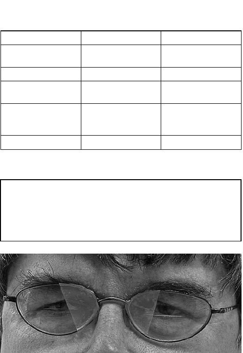
glasses (See Figure 3). He was then instructed to walk
down the hallway. He seemed to be more confident
moving through doorways and past those walking by
him. He also reported feeling better. e BNO was
then added to his home therapy. He was instructed to
ride in his car as a passenger with the BNO in place
to see how he felt during this activity. If he was able to
function comfortably in the car, it was recommended
that he continue to utilize the BNO for the remainder
of the week. If the binasals hindered his ability to
drive, he was instructed to remove the tape.
e following week the patient reported that the
BNO helped his mobility, but he did not feel comfort-
able driving with them on. After his therapy session, we
tried different placements of the BNO until we found
the occlusive area that felt the most comfortable and
maximized the mobility improvement. Optometric
vision therapy was continued for two more weeks
with the BNO in place. Due to scheduling issues, he
discontinued therapy but returned three months later.
At that visit, the binasal occlusion was adjusted and it
was suggested that therapy resume with an emphasis
on home activities. e patient, unfortunately did not
follow through with this plan of action. After a six
month absence, he was contacted by phone at which
time the patient stated that he was still wearing the
BNO for most activities except driving. e patient
noted that he feels much more comfortable walking
with the binasals on, but that his peripheral vision is
restricted while driving. He will continue to be seen
yearly as he did not wish to continue vision therapy
due to transportation difficulties.
DISCUSSION
Binasal Occlusion
Optometrists have treated patients with binasal
occlusion for over fifty years, and have used binasal
occlusion in the treatment of several functional vision
conditions (See Table 2). Numerous case reports
have been written regarding the use of BNO, but the
neurophysiology behind this treatment option has
not been fully documented in the literature.
How does binasal occlusion lead to improved visual
function for TBI patients like the one documented
in the case report? Greenwald used “defocusing” to
describe a “forceful change” in the patient’s habitual
way of handling his visual problem. Binasal occlusion
acts as a “persistent change in the patient’s central visual
space forcing [them]” to alter the way they handle the
world.
1
Binasal occlusion also makes the patient more
dependent on peripheral clues.
5
It reduces confusion
and helps stabilize the image, helping the patient to
understand where they are in their personal visual
space and how to adjust to changes regarding that
space.
Gallop
12
reported on the use of binasal occlusion
for an adult with cerebral palsy. He proposed that
BNO was successful for several reasons. First, by
occluding the middle of the binocular nasal field
there is an effect on binocular integration. Because
this portion of the visual field overlaps, it is an area of
intense demand which requires the highest degree of
binocular integration to be efficient and comfortable.
When this process is changed by acquired brain injury
or strabismus, the integration of this portion of visual
space may be the most difficult when trying to produce
single, clear, comfortable, and stable binocular vision.
If the input from this area is modified, it may serve
to relieve visual stress. is alleviates the confusion of
trying to organize this portion of space and allows the
patient to process information from the peripheral
areas. In the end, this may reduce the patient’s
perception of overall visual stress.
12
Figure 3: Initial placement of the binasal occlusion.
Table 1. Optometric Goals and Sample of Vision Therapy
Activities
Optometric Goal Office Therapy Home Therapy
Improved fixation Pointer in the straw
Brock string
Pointer in the straw
Brock string
Improved pursuits Marsden ball Thumb pursuits
Improved saccades Wayne saccadic
fixator
Saccadic fixation
sheet
Improved
peripheral
awareness
Wayne saccadic
fixator (central to
periphery program)
Fixation Worksheet
VO Star symmetry Line walk
Table 2. Conditions Treated with Binasal Occlusion
3,10,11
Amblyopia•
Anomalous •
Correspondence
Post-Trauma •
Vision Syndrome
Esotropia•
Vertical •
Imbalances
Visually “Tight” •
Patients
Esophoria •
Asthenopia•
Highly Central •
Patients

Volume 40/Number 1/2009
49
His second point was that binasal occlusion allows
the temporal visual fields to become more stimulated
causing greater awareness of the environment. If an
incident causes the patient to shut down peripheral
awareness, it reduces the information crucial to
allowing the patient to feel secure about his relationship
between himself and the world around him. is
increases stress, which can be manifested in other
areas of the body. is stress may reduce peripheral
awareness, creating a vicious cycle.
12
e final point Gallup proposed was that binasal
occlusion provides a visual reference point which
helps to steady visual-spatial perception. Binasals may
act as a constant and consistent reminder, even
if not consciously noticed, interjected between the
patient and the visual environment to help organize
visual input.
12
Traumatic Brain Injury
Patients who have suffered from a traumatic brain
injury will frequently experience trouble with balance,
spatial orientation, coordination, cognitive function,
and speech.
13
Often TBI patients also experience
symptoms such as double vision, apparent movement
of print or stationary objects like walls or floors, eye
strain, visual fatigue, light sensitivity, and headaches.
Visual symptoms are one of the most common
problems following a TBI. Visual dysfunctions may
also manifest as anxiety and panic disorders.
13
Several
articles written by Padula have documented what he
termed “Post-Trauma Vision Syndrome” (PTVS).
14, 15
(See Table 3) Patients not properly treated for PTVS
can experience symptoms for many years following a
neurological event. Padula suggested that treatment
of PTVS include binasal occlusion in conjunction
with low amounts of base-in prism and optometric
vision therapy.
16
Padula and Argyris have noted that many of the
clinical findings associated with TBI are caused by
a dysfunction of the ambient (magnocellular) visual
process. Neurological events like TBI and multiple
sclerosis, and conditions like cerebral palsy can
cause the ambient visual processing system to lose
the ability to match information with other parts of
the sensory-motor feedback loop. Even a whiplash
can cause significant problems at the level of the
midbrain.
13
In most cases, damage from this type
of injury cannot be seen on a CT or MRI, but the
patient will experience symptoms.
13
Dysfunctional
ambient vision can cause patients to have problems
while they are moving through a crowd. e normal
functioning ambient system usually stabilizes images
of the peripheral retina, but in a patient who has had
TBI, this situation may cause complaints of vertigo.
16
e patient may not be able to cope and will avoid
situations where moving through groups of people is
necessary.
Padula, Argyris, and Ray have used visual evoked
potentials (VEP) to study the damage occurring
in the midbrain. In their research two groups were
given binocular visual evoked cross-pattern reversal
P-100 evaluations while wearing their best distance
correction. e experimental and control groups were
given the VEP test. Testing was then repeated with
the subjects in the experimental group while wearing
binasal occlusion and base-in prism. e results of
these tests showed an increase in the amplitude of
the VEP in the experimental group while they were
wearing the binasal occlusion and base-in prism. e
authors suggested that by producing a change in the
ambient vision process via BNO and prisms, the
binocular cortical cells increased in effectiveness. e
patients also objectively noticed that while wearing
the binasal/prism glasses, they saw less movement
of the letters on the chart and for some the diplopia
disappeared completely.
17
Binasal occlusion may not be an appropriate
treatment option for all TBI patients. Although
Jacques was not addressing binasal occlusion as a
treatment for brain injury, some of his cautionary
statements apply to these patients as well. He
indicated that some patients may experience feelings
of discomfort as they cannot or will not function with
any type of treatment that obstructs the use of their
central field. Forcing patients to cope with increased
peripheral awareness may bring a great deal of
distraction into their visual system.
11
Many patients
Table 3. Post-Trauma Vision Syndrome (PTVS)
14,15
Characteristics Symptoms
Exotropia Diplopia
Exophoria Objects Appear to Move
Accommodative Dysfunction Memory Problems
Convergence Insufficiency Staring Behavior
Poor Tracking Ability Asthenopic
Sensitivity to Light Fatigue
Low Blink Rate Difficulty Reading
Spatial Disorientation Irritability
Balance and Postural
Difficulties
Inability to Follow Sequential
Instructions
Visual Memory Problems

50
Optometry & Vision Development
with TBI may benefit from smaller sections of binasal
occlusion. Aiming for areas nasal to the limbus rather
than covering the pupil is recommended for optimal
success in treating these patients.
4
Conclusion
e value of binasal occlusion in treating patients
with brain injury has been demonstrated on an
individual basis. After TBI, many associated visual
dysfunctions may be related to the ambient system’s
inability to process and organize spatial information
which in turn reduces the focal system’s ability to
process incoming information. When BNO is used,
the patient is provided an opportunity to experience
a change which creates the potential for improved
symptomology and behavior.
5
is simple procedure
can be utilized alone as a form of effective therapy or
in conjunction with active therapy. It is inexpensive,
easy to apply, and readily available to all practitioners.
e success reported on a case by case basis has
encouraged clinicians to try this technique on similar
patients. Additional case reports, research and
controlled clinical trials are necessary to validate the
use of binasal occlusion. By utilizing BNO as a part
of the therapeutic regimen, we can change those with
traumatic brain injury so that they can interact with
their world in an effective and productive manner.
References
Greenwald I. Re-evaluation of binasal occlusion. Optom Weekly 1974 Jan 1.
24; 65(4):21-2.
Tassinari J. Binasal occlusion. J Behav Optom 1990;1(1):16-21.2.
Griffin JR. Binocular anomalies: diagnosis and vision therapy. Woburn, 3.
MA: Elsevier Science, 2002:325-6.
Personal conversation with Montecalvo B. and Knueppel K. at Invitational 4.
Lens Symposium in Tahlequah, OK on September 13, 2008.
Greenwald I. Effective strabismus therapy. Santa Ana, CA: OEP, 1979. 5.
Maples WC, Bither M. Efficacy of vision therapy as assessed by the COVD 6.
Quality of Life checklist. Optometry 2002;73(8):492-8.
Maples WC, Atchley J and Ficklin T. Northeastern State University College 7.
of Optometry’s oculomotor norms. J Behav Optom 1992;3(6):143-150.
VO Star by John Streff. Paper and Presentation at the Invitational Lens 8.
Symposium in Tahlequah, OK on September 12, 2008.
Padula W. A behavioral vision approach for persons with physical disabilities. 9.
Santa Ana, CA: OEP, 1988.
VanHoy M. Binasal occlusion. First annual international lens symposium. 10.
Columbus, OH. 2001.
Cox JL. Binasal occlusion. Vision erapy 1996;38(1):56-61.11.
Gallop S. A variation on the use of binasal occlusion: a case study. J Behav 12.
Optom 1998;7(6):31-5.
Padula W, Argyris S. Post-trauma vision syndrome: part I. Centre for 13.
Neuro Skills http://www.neuroskills.com/tbi/vision1.shtml. Last accessed
08/25/2008.
Padula WV, Shapiro JB. Head injury causing post trauma vision syndrome. 14.
N Engl J Optom 1988; Dec: 16-22.
omas JA. e neuro-biochemical basis of post-trauma vision syndrome. 15.
Centre for Neuro Skills http://www.neuroskills.com/tbi/vision3.shtml. Last
accessed 08/25/2008.
Padula W, Argyris S. Post-trauma vision syndrome: part II. Centre for 16.
Neuro Skills
. Last accessed 08/25/2008.17.
Padula W, Argyris S, Ray J. Visual evoked potentials (VEP) evaluating 18.
treatment for post-trauma vision syndrome (PTVS) in patients with
traumatic brain injury. Brain Injury 1994;8(2):125-133.
Materials
A. Bernell (4016 N Home Street, Mishawaka, IN, 46545 or www.bernell.
com).
Did you know?
Optometry & Vision Development (OVD)
is the official quarterly publication of the
College of Optometrists in Vision Development.
COVD is pleased to announce that all issues
of the journal, including the most recent
publications, are available to download by
the public.
e COVD website is the place to:
+ Search for articles
+ Browse past issues
+ Review research and literature
+ View the instructions to authors
+ Order back issues
View Optometry & Vision Development online at
www.covd.org
